刘江 1吴宝林 2朱万招 2刘洁 3王彤 4耿毛毛 2白莉 5刘毅 4
1榆 林 市 第 一 医 院 烧 伤 整 形 外 科 ,榆 林 719000;2 宁 夏 医 科 大 学 临 床 医 学 院 ,银 川750000;3 解放军联勤保障部队第 940 医院烧伤整形外科,兰州 730050;4 兰州大学第二医院烧伤整形与创面修复外科,兰州 730030;5 榆林市中医医院重症医学科,榆林719000通信作者:刘毅,Email:该Email地址已收到反垃圾邮件插件保护。要显示它您需要在浏览器中启用JavaScript。
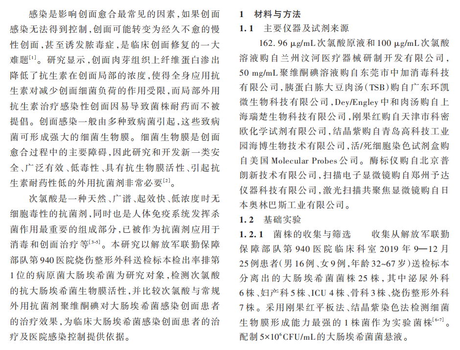
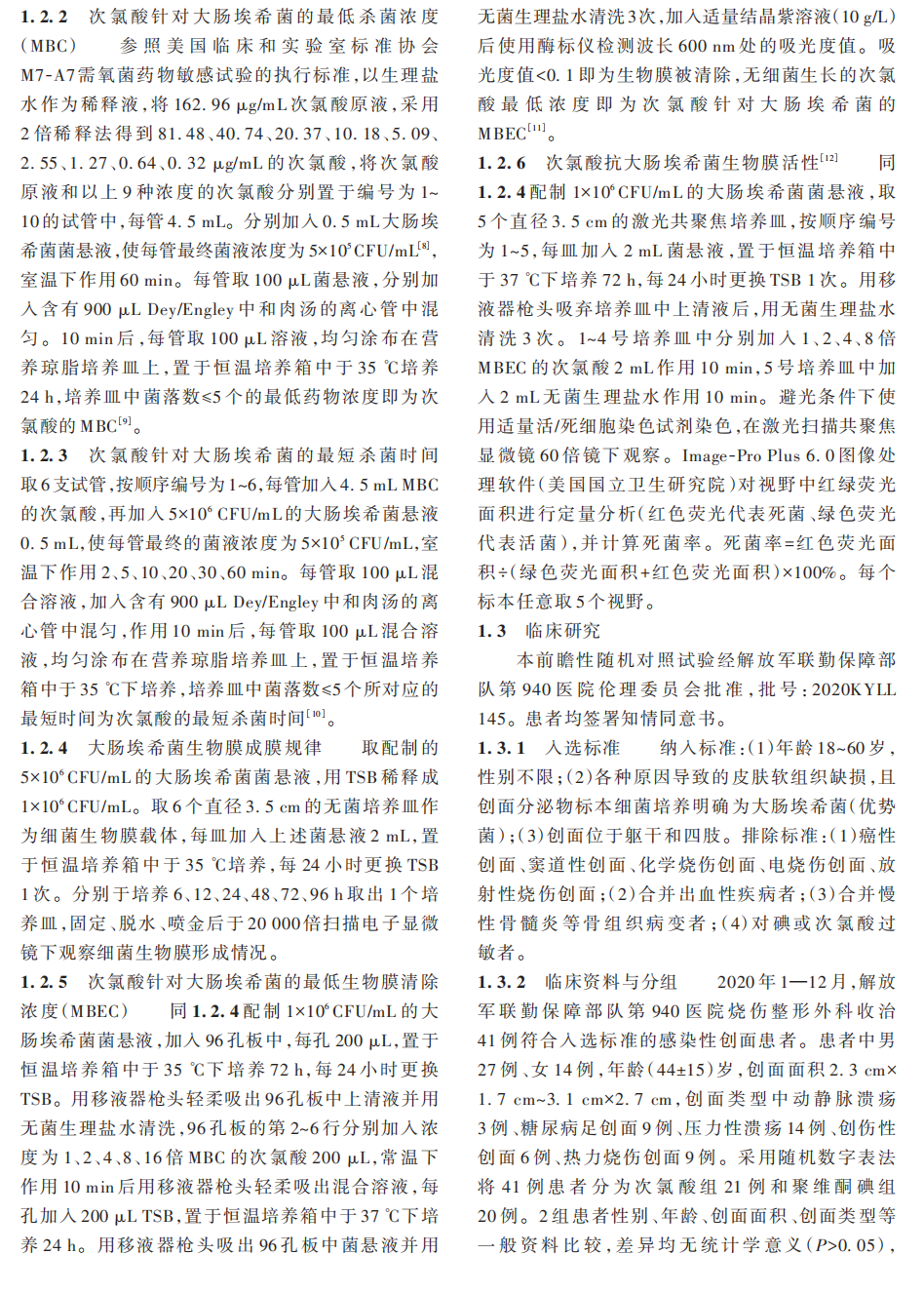
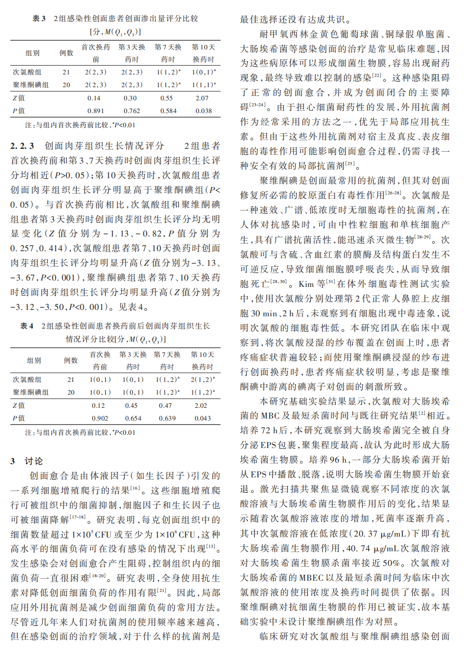
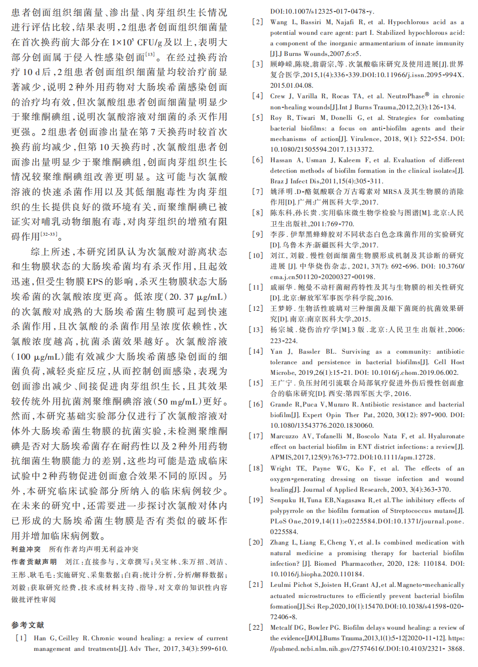
【摘要】目的 探讨次氯酸对大肠埃希菌生物膜的作用及其对大肠埃希菌感染创面的临床疗效。方法 收集从解放军联勤保障部队第 940 医院 5 个临床科室 2019 年 9—12 月 25 例患者(男16 例、女 9 例,年龄 32~67 岁)送检标本分离出的大肠埃希菌菌株中细菌生物膜形成能力最强的 1 株菌进 行 实 验 研 究 。 将 大 肠 埃 希 菌 分 别 与 162.96、81.48、40.74、20.37、10.18、5.09、2.55、1.27、0.64、0.32 μg/mL 的次氯酸共培养,筛选次氯酸最低杀菌浓度(MBC);将大肠埃希菌与筛选的 MBC 次氯酸分别作用 2、5、10、20、30、60 min,筛选次氯酸的最短杀菌时间。分别于培养 6、12、24、48、72、96 h,采用扫描电子显微镜观察大肠埃希菌生物膜形成情况。大肠埃希菌培养 72 h 后,分别加入 1、2、4、8、16 倍MBC的次氯酸,筛选次氯酸对大肠埃希菌的最低生物膜清除浓度(MBEC)。于大肠埃希菌中分别加入 1、2、4、8 倍 MBEC 的次氯酸及无菌生理盐水,作用 10 min 后,采用活/死细菌染色试剂盒检测活、死细胞数,并计算死菌率(样本数为 5)。2020 年 1—12 月,解放军联勤保障部队第 940 医院烧伤整形外科收治 41 例符合入选标准的感染创面患者,对其进行前瞻性随机对照试验。采用随机数字表法将患者分为次氯酸组 21 例[男 13 例、女 8 例,年龄(46±14)岁]和聚维酮碘组 20 例[男 14 例、女 6 例,年龄(45±19)岁]。2 组患者分别用 100 μg/mL 次氯酸、50 mg/mL 聚维酮碘溶液浸透的无菌纱布湿敷,每天换药 1 次。首次换药前、第 10 天换药时,取创面及创缘组织,采用琼脂培养法培养细菌并定量分析组织细菌量。首次换药前和第 3、7、10 天换药时,肉眼观察创面渗出量和肉芽组织生长情况并评分。对数据行单因素方差分析、Dunnett-t检验、独立样本 t检验、Mann-Whitney U 检验、Wilcoxon 符号秩检验、χ2检验或 Fisher 确切概率法检验。结果 次氯酸对大肠埃希菌的 MBC 为 10.18 μg/mL,MBC 的次氯酸对大肠埃希菌的最短杀菌时间为 2 min。培养 6、12 h,大肠埃希菌处于完全游离状态;随着培养时间的延长,大肠埃希菌逐渐聚集、黏附,至培养 72 h 形成成熟的生物膜。次氯酸对大肠埃希菌的MBEC 为 20.36 μg/mL。与 1、2、4、8 倍 MBEC 的次氯酸作用 10 min 后,大肠埃希菌死菌率均明显高于与无菌生理盐水作用 10 min 后(t 值分别为 6.11、25.04、28.90、40.74,P<0.01)。第 10 天换药时,次氯酸组 患 者 创 面 组 织 细 菌 量 为 2.61(2.20,3.30)×104集 落 形 成 单 位(CFU)/g,明 显 少 于 聚 维 酮 碘 组 的4.77(2.18,12.48)×104CFU/g(Z=2.06,P<0.05);次氯酸组和聚维酮碘组患者创面组织细菌量均明显少于首次换药前的 2.97(2.90,3.04)×106、2.97(1.90,7.95)×106 CFU/g(Z 值分别为 4.02、3.92,P<0.01)。第10 天换药时,次氯酸组患者创面渗出量评分明显低于聚维酮碘组(Z=2.07,P<0.05)。与首次换药前比较,次氯酸组患者第 7、10 天换药时创面渗出量评分均明显降低(Z 值分别为−3.99、−4.12,P<0.01),聚106、2.97(1.90,7.95)×106维酮碘组患者第 7、10 天换药时创面渗出量评分均明显降低(Z 值分别为−3.54、−3.93,P<0.01)。第10 天换药时,次氯酸组患者创面肉芽组织生长评分明显高于聚维酮碘组(Z=2.02,P<0.05)。与首次换药前比较,次氯酸组患者第 7、10 天换药时创面肉芽组织生长评分均明显升高(Z 值分别为−3.13、−3.67,P<0.01),聚维酮碘组患者第 7、10 天换药时创面肉芽组织生长评分均明显升高(Z 值分别为−3.12、−3.50,P<0.01)。结论 次氯酸对游离状态和生物膜状态的大肠埃希菌均有杀灭作用,低浓度的次氯酸对成熟的大肠埃希菌生物膜可起到快速杀菌作用,且次氯酸浓度越高,杀菌效果越好。100 μg/mL 次氯酸能有效减少患者大肠埃希菌感染创面的细菌负荷,表现为创面渗出的减少、间接促进肉芽组织生长,较传统外用抗菌剂聚维酮碘疗效更好。
【关键词】 次氯酸; 大肠杆菌; 生物膜; 感染性创面
Effect of hypochloric acid on Escherichia coli biofilm and the clinical efficacy of hypochloric acid for wounds with Escherichia coli infection
Liu Jiang1 , Wu Baolin2 , Zhu Wanzhao2 , Liu Jie3 , Wang Tong4 , Geng Maomao2 , Bai Li5 , Liu Yi4
1Department of Burns and Plastic Surgery, the First Hospital of Yulin, Yulin 719000, China; 2Clinical Medical College, Ningxia Medical University, Yinchuan 750000, China; 3Department of Burns and Plastic Surgery, the 940th Hospital of the Joint Logistic Support Force of PLA, Lanzhou 730050, China; 4Department of Burns andPlastic Surgery & Wound Repair Surgery, Lanzhou University Second Hospital, Lanzhou 730030, China; 5Intensive Care Unit, Traditional Chinese Medicine Hospital of Yulin, Yulin 719000, China Corresponding author: Liu Yi, Email: 该Email地址已收到反垃圾邮件插件保护。要显示它您需要在浏览器中启用JavaScript。
【Abstract】 Objective To investigate the effect of hypochloric acid on Escherichia coli biofilm and the clinical efficacy of hypochloric acid for wounds with Escherichia coli infection. Methods One strain of Escherichia coli with the strongest bacterial biofilm forming ability among the strains isolated from specimens in 25 patients (16 males and 9 females, aged 32−67 years) from five clinical departments of the 940th Hospital of the Joint Logistic Support Force was collected for the experimental study from September to December 2019. The Escherichia coli was cultured with hypochloric acid at 162.96, 81.48, 40.74, 20.37,10.18, 5.09, 2.55, 1.27, 0.64, and 0.32 μg/mL respectively to screen the minimum bactericidal concentration(MBC) of hypochloric acid. The Escherichia coli was cultured with hypochloric acid at the screened MBC for2, 5, 10, 20, 30, and 60 min respectively to screen the shortest bactericidal time of hypochloric acid. The biofilm formation of Escherichia coli was observed by scanning electron microscopy at 6, 12, 24, 48, 72, and 96 h of incubation, respectively. After 72 h of culture, hypochloric acid at 1, 2, 4, 8, and 16 times of MBC was respectively added to Escherichia coli to screen the minimum biofilm eradicate concentration (MBEC) of hypochloric acid against Escherichia coli. After hypochloric acid at 1, 2, 4, and 8 times of MBEC and sterile saline were respectively added to Escherichia coli for 10 min, the live/dead bacterial staining kit was used to detect the number of live and dead cells, with the rate of dead bacteria calculated (the number of samples was 5). From January to December 2020, 41 patients with infectious wounds meeting the inclusion criteria and admitted to the Department of Burns and Plastic Surgery of the 940thHospital of Joint Logistic Support Force of PLA were included into the prospective randomized controlled trial. The patients were divided into hypochloric acid group with 21 patients (13 males and 8 females, aged (46±14) years) and povidone iodine group with 20 patients (14 males and 6 females, aged (45±19) years) according to the random number table. Patients in the 2 groups were respectively dressed with sterile gauze soaked with hypochloric acid of 100 μg/mL and povidone iodine solution of 50 mg/mL with the dressings changed daily. Before the first dressing change and on the 10th day of dressing change, tissue was taken from the wound and margin of the wound for culturing bacteria by agar culture method and quantifying the number of bacteria. The amount of wound exudate and granulation tissue growth were observed visually and scored before the first dressing change and on the 3rd, 7th, and 10th days of dressing change. Data were statistically analyzed with one-way analysis of variance, Dunnett-t test, independent sample t test, Mann-Whitney U test, Wilcoxon signed-rank test, chi-square test, or Fisher's exact probability test. Results The MBC of hypochloric acid against Escherichia coli was 10.18 μg/mL, and the shortest bactericidal time of hypochloric acid with MBC against Escherichia coli was 2 min. Escherichia coli was in a completely free state after 6 and 12 h of culture and gradually aggregated and adhered with the extension of culture time, forming a mature biofilm at 72 h of culture. The MBEC of hypochloric acid against Escherichia coli was 20.36 μg/mL. The Escherichia coli mortality rates after incubation with hypochloric acid at 1, 2, 4, and 8 times of MBEC for 10 min were significantly higher than that after incubation with sterile saline (with t values of 6.11, 25.04, 28.90, and 40.74, respectively, P<0.01). The amount of bacteria in the wound tissue of patients in hypochloric acid group on the 10th day of dressing change was 2.61 (2.20, 3.30)×104colony forming unit (CFU)/g, significantly less than 4.77 (2.18, 12.48)×10
4CFU/g in povidone iodine group (Z=2.06, P<0.05). The amounts of bacteria in the wound tissue of patients in hypochloric acid group and povidone iodine group on the 10th day of dressing change were significantly less than 2.97 (2.90, 3.04)×106 and 2.97 (1.90, 7.95)×106 CFU/g before the first dressing change (with Z values of 4.02 and 3.92, respectively, P<0.01). The score of wound exudate amount of patients in hypochloric acid group on the 10th day of dressing change was significantly lower than that in povidone iodine group (Z=2.07, P<0.05). Compared with those before the first dressing change, the scores of wound exudate amount of patients in hypochloric acid group on the 7th and 10th days of dressing change were significantly decreased (with Z values of −3.99 and −4.12, respectively, P<0.01), and the scores of wound exudate amount of patients in povidone iodine group on the 7thand 10th days of dressing change were significantly decreased (with Z values of −3.54 and −3.93, respectively, P<0.01). The score of wound granulation tissue growth of patients in hypochloric acid group on the 10th day of dressing change was significantly higher than that in povidone iodine group (Z=2.02, P<0.05). Compared with those before the first dressing change, the scores of wound granulation tissue growth of patients in hypochloric acid group on the 7thand 10th days of dressing change were significantly increased (with Z values of − 3.13 and − 3.67, respectively, P<0.01), and the scores of wound granulation tissue growth of patients in povidone iodine group on the 7th and 10th days of dressing change were significantly increased (with Z values of −3.12 and −3.50, respectively, P<0.01). Conclusions Hypochloric acid can kill Escherichia coli both in free and biofilm status. Hypochloric acid at a low concentration shows a rapid bactericidal effect on mature Escherichia coli biofilm, and the higher the concentration of hypochloric acid, the better the bactericidal effect. The hypochloric acid of 100 μg/mL is effective in reducing the bacterial load on wounds with Escherichia coli infection in patients, as evidenced by a reduction in wound exudate and indirect promotion of granulation tissue growth, which is more effective than povidone iodine, the traditional topical antimicrobial agent.
【Key words】 Hypochloric acid; Escherichia coli; Biofilm; Infectious wounds
