
Welcome to the next instalment of 'Wound detectives'. Joy Tickle shares a real-world case presentation and asks whether you can solve the case. What do you think is the cause of the wound, what tests would you order to confirm your diagnosis and what treatment would you provide?
JOY TICKLE Tissue Viability Nurse Consultant
When I was asked to write a further episode of the Wound Detectives I sat and reflected on my 27 years' experience as a Tissue Viability Specialist. This took me to a memory I have held for many years and a patient I met when I was in my first year as a tissue viability nurse.
John was referred to me by the community nursing team with a diagnosis of a long standing chronic venous leg ulcer (VLU) that would reduce in size and almost heal and
Along with the community nurse I visited John at home. He lived alone and, although he found it difficult to leave his home, was independent with meeting his everyday activities and needs. It is important before assessing any patients wound that we talk with them to discuss key factors such as how the wound occurred, longevity of the wound previous treatments and interventions and the impact of the wound upon their quality of life then deteriorate and breakdown further.
This can offer valuable and rich information to assist the clinician in ascertaining a true diagnosis and factors/ reasons why the ulceration is non-healing. It can also assist in identifying the impact for the patients living with a non-healing wound so that actions may be undertaken to improve any negative aspects of this. John reported that the wound was painful and sometimes “smelled “and that he had had the leg ulcer for over 20 years and on occasions the ulcer would close but then reopen a week or so later.
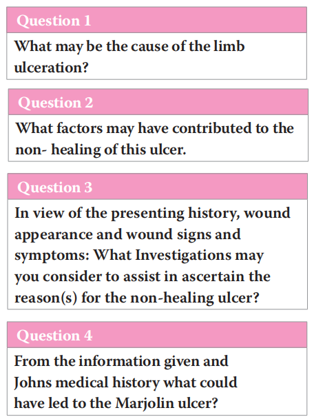
Question 1. What may be the cause of the limb ulceration? Answer: The possible causes may be :
�Venous disease
�Arterial disease
�Previous trauma injury
�Chronic skin/wound infection
�Malignancy
�Inflammatory ulceration
�Dermatological condition.
Question 2. What factors may have contributed to the non-healing of this ulcer.
Answer:
�Wound Infection/bone infection
�Reduced arterial blood supply
�Inadequate compression therapy/ no compression therapy to assist venous return
�Ineffective wound bed preparation and wound management
�Unhealthy periwound/surrounding skin
�Underlying comorbidities such as diabetes, autoimmune disease
�Medication that affects wound healing such as anticoagulants, immunosuppressive treatments
�Behavioural factors such as patient non-concordance with recommended treatment regimen or health education advice
�Poor nutritional/hydration.
I considered all the above factors and discussed each one with the nurse and John. It was evident that John was extremely concordant with his compression hosiery bandage system that delivered 40mmhg pressure to aid venous return and assist in managing his venous disease (Atkin, 2019). The wound treatment regimen involved evidence-based wound debridement and the use of appropriate primary dressings to facilitate wound healing and autolysis and an effective secondary dressing to maintain wound bed moisture balance.
Despite this John’s wound often became malodourous and this caused him significant embarrassment and reluctance to allow visitors to his home. The nurse had introduced a charcoal dressing to assist with the malodour.
From clinical appearance John’s limb showed signs and symptoms of venous disease. Haemosiderin staining and ankle flare were present, and the ulcer was on the lower gaiter region of his limb a common location for venous ulcers to occur (Atkin, 2019). The wound edges were flat there was periwound maceration and the granulation tissue bled easily on dressing change.
Question 3. In view of the presenting history, wound appearance and wound signs and symptoms: What Investigations may you consider to assist in ascertain the reason(s) for the non-healing ulcer?
Answer:
�Repeat vascular studies to assess both venous and arterial blood supply to the limb
�Xray/bone scan to rule out infection to the bone (osteomyelitis)
�Tissue/exudate sample for microbiology (increased accuracy for ascertaining wound microbiology)
�Tissue biopsy for histology to eliminate other aetiologies such as vasculitis, pyoderma gangrenosum, malignancy
�Blood studies to diagnose and address any disorders such as anaemia, malnutrition, or comorbidities such as diabetes.
John underwent several tests and investigations. The arterial supply to his limb was satisfactory and as previously diagnosed he had venous disease. The microbiology results of the swab and exudate sample did not identify any harmful microorganisms other than natural skin and wound flora. However, the x-ray of his limb did identify bone density changes and evidence of osteomyelitis.
Recognised good practice for ulcers that do not respond to treatment and are chronic in nature, is to take a tissue sample. (Pekarek et al, 2011). As I am trained to undertake tissue biopsy sampling, I arranged for John to attend the tissue viability clinic the following week where I took several tissue samples for histology. At this point from the wound appearance, I initially thought that it would be unlikely that malignancy was present. The blood results did indicate John was anaemic and investigations were arranged by the GP to investigate further, and iron supplements commenced.
I reviewed John two weeks later at home at this point the GP and I had received the histology results which identified noninfiltrating, well-differentiated squamous cell carcinoma consistent with a Marjolin ulcer. This was shocking news not only to John but also to the nurse who had been caring for him for many years and for myself. John was obviously distressed by this diagnosis and further advice and ongoing support was given.
Marjolin ulcers are an overlooked entity. The name originates from the man that recognised this condition in 1828, a French surgeon Marjolin JN (Kamran et al, 2016). It is a cutaneous malignancy that can occur on previously injured skin, longstanding scar tissue (commonly burn scars) and other chronic inflammatory aetiologies such as traumatic wounds, venous leg ulcers, osteomyelitis, pressure ulcers, radiation dermatitis (Kamran et al, 2016). They can also develop from amputation scars, skin grafts vaccination scars and stings/bites (Watson, 2021).
Marjolijn’s ulcers occur in all age groups, sexes and races and ethnicities. The average period from the time of the initial wound to the discovery of the malignancy is between 30 and 35 years (Iqbal et al, 2015).
The pathophysiology has not been completely elucidated but there are two main theories.
1. Injury and scar formation lead to destruction of the local blood and lymphatic vessels, making the area an immune-privileged site. This protects the scar from anti-tumour antibodies and permits the transformation and malignant degeneration of the skin (Pekerek, 2011; Junad, 2017).
2. Long-term irritation, chronic inflammation, or trauma to the area causes skin cells to constantly repair themselves. During this renewal process, some skin cells become cancerous (Watson, 2021). Presenting signs of this type of wound are often a malodourous wound, over granulation/bleeding tissue, extreme pain, and non-healing (Saha, 2021).
Question 4. From the information given and John's medical history what could have led to the Marjolin ulcer?
Answer:
�Chronic venous ulcer
�Bone infection (osteomyelitis)
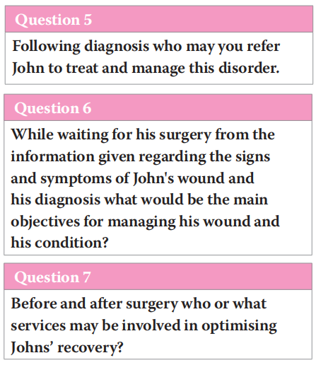
Question 5. Following diagnosis who may you refer John to treat and manage this disorder.
Answer:
�GP
�Dermatologist
�Vascular consultant
�Burns/plastic team
�Oncologist
�Counselling service.
Initially John was referred to the vascular/ surgical team who undertook further investigations this included MRI imaging to assess the degree of soft tissue and bone involvement. Due to the length of time the wound had been present, the aggressive nature of a Marjolin ulcer and its ability to metastasis an MRI scan of John's chest/ abdomen and lymphatics were performed. Thankfully the cancer had not spread to his lymphatic system or other organs however unfortunately metastasis was identified in the limb bone.
The mainstay treatment for a Marjolin ulcer is surgery. A surgeon can use a few different methods to do this, including: Excision. This method involves removing the tumour and a margin of extended tissue surrounding the ulcer.
Mohs surgery. in which a surgeon serves as both surgeon and pathologist in the operating room, in this technique, the tissue is immediately examined after excision to determine whether the margins are clear. (Holgado et al, 2000; Shah et al, 2021).
Amputation for lesions that involve the bone.
If surgery is not possible or refused radiotherapy may be used for palliation for these patients.
Although most Marjolijn’s ulcers are well differentiated lesions are aggressive and carry a poor prognosis. Metastases as in John’s case are found in up to 27% of patients, the recurrence rate after surgical resection is up to 50% and the 5-year survival rate is 43% to 58% (Baskara et al, 2010). Consequently, regular monitoring for at least 3 years is imperative for patients who have or have had a Marjolin ulcer (Onesti et al, 2015).
Due to the presence of metastases to the bone in John’s leg the consultant recommended a below knee amputation to remove the diseased bone and Marjolin ulcer and to assist in trying to prevent further spread of the cancer.
Question 6. While waiting for his surgery from the information given regarding the signs and symptoms of John's wound and his diagnosis what would be the main objectives for managing his wound and his condition?
Answer:
�Atraumatic dressing change
�Comfortable dressing to assist with pain management
�Effective dressing wear time in order to avoid unnecessary dressing changes which may cause him additional pain and distress
�Antimicrobial primary dressing and secondary charcoal dressing to assist with the malodour from the wound and improve his quality of life.
�Effective pain assessment and management
�Support and counselling related to his diagnosis and forth coming surgery.
It is important to remember that for this type of ulcer wound healing/closure is not the main objective as Marjolin ulcers will not heal unless the lesion is removed. The overall objectives are to prevent infection and promote comfort for the patient.
After 3 weeks from initial diagnosis John was admitted to hospital and underwent the planned below knee amputation.
Question 7. Before and after surgery who or what services may be involved in optimising Johns’ recovery?
Answer:
�Community nursing team
�Occupational therapy team
�Physiotherapist
�Orthotic/limb services
�Social services
�Community rehabilitation team
�Patient support group
�Counselling service.
CONCLUSION
Malignant degeneration in wounds and scar tissue can occur. It is essential that wound-care providers and clinicians caring for patients with wounds are aware of different wound aetiologies and able to identify the signs and symptoms of malignant degeneration in wounds. This will ensure early diagnosis that may decrease the risk of tissue destruction and extensive surgical reconstruction and increase the possibility of a good prognosis.
Wounds that are failing to heal or frequently reoccur should be investigated further and appropriate referrals made to the wider multi-disciplinary team.
I hope that this episode of wound detective will support you understanding the importance of further investigations if a wound fails to heal and in recognising and learning about Marjolin ulcers.
REFERENCES
1. Atkin L (2019) Venous Leg Ulceration 1:Identifying Patients who are at risk. Nurs Times [online issue]; 115:6:24–28.
2. Baskara A, Sikka ., Khan F, Sapanara N (2010) Development of a Marjolin's ulcer within 9 months in a plantar pressure ulcer. Eur J Dermatol 20(2):225. https://doi.org/10.1684/ ejd.2010.0850
3. Holgado RD, Ward SC, Suryaprasad SG (2000) Squamous cell carcinoma of the hallux. J Am Podiatr Med Assoc 90(6):309–311. https://doi.org/10.7547/87507315-90-6- 309
4. Iqbal FM, Sinha Y, Jaffe W (2015) Marjolin’s ulcer: a rare entity with a call for early diagnosis. BMJ Case Rep 2015:bcr2014208176. https://doi.org/10.1136/bcr-2014- 208176
5. Junaid F(2017) Marjolin Ulcer https://dermnetnz.org/topics/ marjolin-ulcer Accessed 2/2/2022
6. Kamran K, Giannone,A, MehrabiF (2016) Marjolin’s ulcer complicating a pressure sore: the clock is ticking. Am J Case Rep 17:111–114. https://dx.doi. org/10.12659%2FAJCR.896352
7. Onesti MG, Fino P, Fioramonti P (2015) Ten years of experience in chronic ulcers and malignant transformation. Int Wound J 12(4):447–50. https://doi.org/10.1111/iwj.12134
8. Pekarek B, Buck S, Osher L (2011) A comprehensive review on Marjolin's ulcers: diagnosis and treatment. J Am Col Certif Wound Spec 3(3): 60–4. PubMed Published online 2012 Jul 4. doi: 10.1016/j.jcws.2012.04.001
9. Shah M, Crane J (2021) Marjolin Ulcer .https://www.ncbi. nlm.nih.gov/books/NBK532861/ Accessed 2/2/2022
10. Watson S (2021) Marjolin Ulcers How does it develop? Accessed 2/2/2022) Marjolin Ulcer: Image, Causes, Treatment, and Symptoms (healthline.com)
This article is excerpted from the Wounds UK | Vol 18 | No 1 | 2022 by Wound World.

