
Issue: Volume 53 - Issue 5 - May, 2007
Index: Ostomy Wound Manage. 2007;53(5):30-37.
Login or Register to download PDF
The use of bioprosthetic materials is an evolving strategy in reconstructing soft tissue defects. The use of bioprosthetics to facilitate repair of complex abdominal wall defects has been reported but remains limited. After reviewing published indications and outcomes of using bioprosthetic materials to reconstruct complex abdominal wall defects, the safety and use of two different types of bioprosthetic materials were assessed in two patients requiring repair of their abdominal wall defect.
Literature Review
A literature search was conducted using the PubMed™ search engine and utilizing the following search terms, alone or in combination: Alloderm, Permacol, abdominal wall reconstruction, bioprosthesis, human acellular dermis, and porcine tissue matrix. The entire database since the inception of PubMed™ (including the “old” MEDLINE) through February 2007 was searched. In addition, cross-referenced articles not indexed in PubMed™ were included in this review. Eight case reports and case series, for a total of 137 cases, were identified.1-8
Of the 137 reported cases, the most common indications for the use of dermal-derived materials in this review were reconstruction of abdominal wall defect following pedicle transverse rectus abdominus musculocutaneous (TRAM) flap creation (54, 39%), difficult or recurrent ventral hernia repair with concurrent infection/contamination or high risk of contamination (29, 21%), open abdominal management of a general surgical or trauma patient (25, 18%), large abdominal wall defects following radical oncologic excisions (14, 10%), abdominal wall reconstruction involving active infection and presence of a fistula (eight, 5.8%), and abdominal dehiscence with concurrent abdominal sepsis (seven, 5.1%) (see Table 1).
Reported indications. Bioprosthetic materials have been utilized in breast reconstruction, burn wound management, various hernia repairs, and plastic-reconstructive procedures.2,9-11 The literature suggests that these materials are generally safe and effective. Their use has gradually expanded to include complex abdominal reconstruction indications.1-5,9,12,13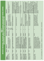
The goal of abdominal wall reconstruction is to restore and maintain abdominal domain.12 This can be a difficult undertaking, especially when the fascial defect is large and underlying viscera are exposed.1 Repair of abdominal wall defects requires closure materials that exhibit certain properties, including the ability to adequately support abdominal structures and easily accommodate a wide variety of defect shapes and sizes.1 The collagen and elastin found both in human and porcine dermal-derived bioprostheses have been shown in clinical or preclinical studies to provide the necessary durability and strength required to prevent hernia recurrence.1,2,4,9 In addition, in one clinical study involving 13 patients, the human dermal-derived graft was found to revascularize within 1 to 4 weeks after implantation.1
Complications. Reported complications associated with the use of human and porcine dermal-based bioprosthetic materials for fascial replacement in 137 patients (see Table 1) during a follow-up period of 6 to 18 months include: wound seroma (18, 13%), skin dehiscence with graft exposure without herniation (six, 4.4%), superficial and deep wound infections (five, 3.6%), hernia recurrence (four, 2.9%), graft failure with dehiscence (two), hematoma (two), enterocutaneous fistula (one), and flap necrosis (one).1-8 Most reported complications are relatively minor and easily treatable. In addition, some reported complications are part of the overall risk of the surgical procedures and not directly related to the bioprosthetic material.
One particular concern regarding the use of human dermal-derived grafts is bulging at the site of bioprosthetic material placement. In this review, bulging was reported in 12 out of 120 patients (10%).1-8 This phenomenon may be due to the fact that human dermal-derived bioprosthetic materials tend to increase their surface area up to 35% following adequate rehydration and insetting with appropriate physiologic tension, a characteristic that is inversely related to the thickness of the material.1 This, in turn, could lead to eventual appearance of laxity or bulging at the site of the bioprosthesis and functionally result in diastasis-like appearance of the abdominal wall, along with accompanying patient discomfort. This phenomenon appears to be less common with porcine-derived material, perhaps due to its greater thickness and the fact that it does not require rehydration before implantation.8
Comparative advantages over synthetic mesh. It has been shown that the use of synthetic mesh (commonly used to repair abdominal defects) in previously irradiated or contaminated tissues greatly increases complication rates.1 In a recent comprehensive review, long-term hernia recurrence rates in all patients were found to be as high as 40% or more for simple suture repair and up to 10% for open mesh repair.14 With more severe complications – eg, enterocutaneous fistula, flap necrosis, wound infection, and bioprosthetic material infection – the incidence of bioprosthetic material complications is comparable if not lower than that found with synthetic mesh repairs.4,11,14
Bioprosthetic materials offer several advantages over synthetic mesh. In terms of durably in providing for abdominal wall closure, both currently available dermal-derived materials appear to provide long-term success rates that are equal to or better than traditional synthetic mesh-based approaches.1,2,14
Although both types of prostheses are associated with local complications such as seroma, hematoma, flap necrosis, and dehiscence, bioprosthetic materials produce minimal abdominal adhesions because they do not tend to stimulate a foreign body reaction.1-4 This reduced inflammatory response, with resulting decrease in adherence to the intestinal wall, is thought to decrease the formation of enterocutaneous fistulae and allows placement of the bioprostheses directly over exposed viscera.1,4,13
Infection. Bioprosthetic materials appear to be less susceptible to infection due to rapid vascularization, facilitating use even when bacterial contamination and enteric soilage are present.1,6 In one study of 54 patients undergoing abdominal wall donor site reconstruction following pedicle TRAM flap procedures following mastectomy, no infectious complications were reported.7 A study1 from the University of Texas MD Anderson Cancer Center reported only one case of infectious complication (an enterocutaneous fistula due to breakdown of an irradiated colonic anastomos located deep within the abdomen and remote from the allograft) in seven of 13 patients who underwent complex reconstructive procedures using the human dermal-derived allograft in the presence of gross intestinal spillage and/or contaminated wounds in the operative field. In another series,2 patients who underwent abdominal wall reconstruction with human-derived allograft experienced no infectious complications, despite the fact that nine of 16 had either a previous hernia site infection, exposure of old prosthetic material, or old mesh extrusion. A similar series8 from the Eisenhower Army Medical Center reported no immediate infectious complications despite the presence of wound contamination in five of nine patients who underwent abdominal wall reconstruction with porcine dermal collagen material (although one case of delayed infection necessitated graft removal at 13 months due to a suture eroding into the bowel). A study3from Baylor College of Medicine reported superficial infections in only two out of 37 patients who underwent abdominal wall closure using the human acellular dermal matrix, both of which were treated with local wound care; no graft loss was reported.
Approved indications. According to manufacturer and insurance industry publications, decellularized human cadaveric dermis is US Food and Drug Administration (FDA)-approved for use in burns and full-thickness wounds and has been used for tissue reconstruction in situations of soft tissue loss.15 Porcine-derived isocyanate cross-linked collagen tissue matrix has received FDA approval for parastomal hernia and abdominal wall defect applications.16,17 Another class of bioprostheses derived from small intestinal submucosa (Surgisis™, Cook Biotech Incorporated, West Lafayette, Ind)18 has been found to lack sufficient strength to support stresses encountered with some ventral hernias or large-area, body-wall repairs18; plus, although not recommended for large ventral hernias or large-area body wall repair, it is not contraindicated for other uses.
Materials utilized in this report were within approved indications for use. Appropriate permissions and consents were obtained.
Case Studies
Case 1. Ms. R, a 46-year-old woman, was admitted with epigastric pain 19 months after a Roux-en-Y gastric bypass procedure and was found to have free intraperitoneal air on computed tomography (CT) imaging. She was septic with evolving multi-organ failure and underwent a prompt exploratory laparotomy that identified a perforation of the gastric remnant with significant enteric leakage. The perforation was repaired without difficulty; however, because of severe soft tissue and bowel edema, it was determined that primary fascial closure would not be possible at that time. The wound remained open and abdominal vacuum-assisted closure (V.A.C. System, KCI Inc., San Antonio, Tex) was applied. She underwent a series of planned washouts with negative pressure wound therapy dressing changes.
After generalized edema improved and multi-system organ failure resolved, Ms. R was taken for definitive abdominal fascial closure using porcine-derived isocyanate cross-linked collagen tissue matrix (Permacol™, Tissue Science Laboratory, Hampshire, UK). Large sheets of porcine tissue matrix were used to reconstruct the fascial defect that measured approximately 30 cm cephalad to caudad and 20 cm side-to-side. The sheets were approximated using a combination of running and interrupted non-absorbable sutures (see Figure 1). Subcutaneous tissue overlying the porcine tissue material was mobilized and the skin edges were sutured together using 2-0 nylon interrupted mattress sutures. A closed-suction drain was placed underneath the cutaneous flap. Postoperatively, Ms. R showed continuous improvement and was discharged 21 days after her abdominal closure. She remained healthy 5 months after discharge.
Case 2. Ms. S, a 54-year-old woman, was seen in the surgical clinic with a left abdominal wall mass. She had been treated abroad for a presumed soft tissue sarcoma approximately 9 months earlier. At that time, she underwent excisional biopsy and a subsequent course of external beam radiation. Results of an ultrasound-guided biopsy performed at one of the authors’ institutions demonstrated spindle cell neoplasm. Computed tomography of her abdomen and pelvis showed enhancing soft tissue masses present within the left abdominal wall – the largest mass measured 7.7 cm x 4.9 cm with two satellite nodules (see Figure 2a,b).
Ms. R underwent en-bloc resection of the abdominal wall masses and adjacent tissue, including excision of four ribs and a portion of the diaphragm in order to obtain oncologically negative margins. Following excision, the left abdominal wall defect measured approximately 20 cm x 15 cm (see Figure 3a,b). In order to reconstruct such a large abdominal wall defect, three sheets of decellularized human cadaveric dermis (AlloDerm™, Lifecell Corporation, Branchburg, NJ) were sutured in place using running 2-0 Prolene sutures (see Figure 3c). A curvilinear incision was made medially and a large myocutaneous rotational flap was created to fill the defect. The skin and subcutaneous tissue were closed over two closed suction drains (see Figure 3d). Ms. R left the hospital on postoperative day 6 without complications. The final pathology identified the mass as a leiomyosarcoma of moderate grade. All margins were free of tumor. She was doing well 20 weeks after discharge with no evidence of recurrence or hernia.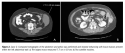
Discussion 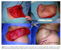
Traditional methods for soft tissue reconstruction include primary closure, closure with synthetic mesh, and closure with myocutaneous flaps.2,4 Dermal-derived bioprosthetic materials, including a decellularized human cadaveric dermis and a porcine-derived isocyanate cross-linked collagen tissue matrix, have been developed to expand available reconstructive options.1,4 In this report of two cases of abdominal wall reconstruction, the results were promising and no apparent complications associated with the use of bioprosthetic materials were observed.
Dermal-derived bioprosthetic materials can be manipulated intraoperatively according to the various shape and size specifications required in abdominal wall defect repairs. These properties are clearly demonstrated in the two cases presented – the patients had large, uneven abdominal wall defects (eg, case 2) located in areas of high stress (both cases). Other cases1demonstrate similar adaptability of bioprosthetic materials in the setting of complicated, irregularly-shaped wounds.
The goal of abdominal wall reconstruction is to restore and maintain the abdominal domain.12 This can be difficult, especially when the fascial defects are large and the underlying viscera are exposed. The use of synthetic mesh in previously irradiated or contaminated tissues increases complication rates; utilizing bioprosthetic material in these cases may help minimize infectious risk and/or adhesions.1,6
The first case in the current report exemplifies the use of bioprosthetic material in an environment of enteric spillage where porcine tissue matrix material is placed directly over the exposed bowel. The second case involves tissue subjected to external beam radiation before radical resection, resulting in a defect where human dermal-derived material was thought to provide the best option. The use of bioprosthetic material allowed successful closure of both wounds without multiple procedures and with no complications noted in the follow-up period.
Given the overall versatility and characteristics of human and porcine dermal-derived grafts, clinical indications for their use are largely interchangeable. In the authors’ experience, the most important determinant for bioprosthetic material utilization was the size of the defect relative to the size and thickness of available prosthetic pieces. The size of the largest available porcine dermal prosthesis (20 cm x 30 cm) renders it better suited for larger defects. In addition, the “bulging” phenomenon of bioprosthetic material following implantation may be related to expansile properties of the implanted graft as well as its thickness; hence, both size and thickness of the material should be considered in the context of each individual wound.
Techniques to improve outcome with bioprosthetic grafts proposed by Butler et al1 deserve consideration. They include use of 1) thicker or extra-thick bioprosthetic material, 2) appropriate technique for joining of multiple sheets, 3) inlay technique with maximal fascia overlap for abdominal wall defects and onlay technique for chest wall defects, and 4) seroma prevention strategies utilizing suction-drainage catheters, quilting sutures, and compression garments.
Although existing literature suggests that the use of human- and porcine-derived prosthesis for complex tissue reconstruction is safe, effective, and may be associated with low recurrence and postoperative infectious rates, prospective, randomized trials comparing this methodology to existing reconstruction techniques have not been conducted.
Conclusion
Bioprosthetic materials are changing the paradigm of abdominal wall reconstruction. In the presence of irradiated and/or contaminated tissues, these materials provide new options for definitive abdominal wall repair. Based on the two cases presented and the review of current literature series and reports, it appears that bioprosthetic materials are safe and effective when used for repair of complex abdominal wall defects. The indications for their use are likely to expand, underscoring the need for prospective, randomized comparison studies to ascertain the efficacy, applicability, indications, risks, and benefits of dermal-derived bioprostheses as compared to other abdominal wall reconstruction methods.