
伤口世界
电子邮件地址: 该Email地址已收到反垃圾邮件插件保护。要显示它您需要在浏览器中启用JavaScript。

- 星期四, 14 11月 2019
朱庆棠
擅长于利用显微外科的独特优势治疗肢体创伤及其并发症、后遗症,如骨折、骨不连、骨坏死、骨髓炎和骨折畸形愈合等的处理;神经、血管、肌肉、肌腱、韧带和皮肤等软组织损伤和缺损的修复;肢体创伤后遗症的治疗、外形与功能的重建。对手与上肢疾患的外科治疗也有较丰富的经验。


- 星期二, 12 11月 2019
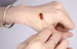
自己怎么做才能使伤口缝合后长得好, 疤痕小?
原创: 白衣山猫 白衣山猫
不小心受伤了,经过医生缝合后,回到了家里,许多人最关心的问题是:伤口为什么要缝合?我该怎么来护理伤口?怎么样才能避免伤口感染?怎么样让伤口长得更好?什么时候才能拆线?怎么样才能够使得我的伤口疤痕最小?今天,我来回答这个问题。

- 星期二, 12 11月 2019
没有长不好的伤口。即使有,那一定不是你的。
原创: 郭铭川 铭医铭言
伤口液化的问题,在妇产科经常会遇到。为啥?因为咱们科“肥”啊!
伤口之所以会液化,是因为皮下脂肪的问题。没有皮下脂肪的人,相对没那么容易出现脂肪液化,但是作为一名风韵的女子,怎么能没有皮下脂肪呢?妇产科就是这样一个风韵十足的科室,伤口脂肪液化自然会多一些。

- 星期二, 12 11月 2019
伤口护士的角色和作用
Matthew 慢伤前沿
为了满足现代专业伤口管理的要求,伤口管理护士(Wound care nurse)这一角色在近年来获得了快速发展和认可。纵观国际社会,这一角色在不同国家和地区具有不同的称谓和岗位。尽管所有的职位都有一些共同的任务和职责,但围绕着伤口护士这一角色仍然存在知识的断层。今天分享的这片文章通过大量文献的回顾,对当前伤口护士的执业环境、职责范围和发挥的影响(或作用)进行了总结。虽然国际上对于伤口护士所需的能力水平、教育和资质等并没有共识。但有证据显示伤口护士改善了伤口的愈合时间和降低了压力性损伤的发生率。
