
伤口世界
- 星期三, 22 7月 2026
An Acid-Oxidising Solution Containing Hypochlorous Acid Reduces Staphylococcus aureus and Improves Bacterial Diversity in Epidermolysis Bullosa Wounds
Xiaolong A. Zhou1 | Michael B. Burns2 | Ziyou Ren1 | Elise Stagaman2 | Stefan J. Green3 | Lok Yiu Ashley Wu3 |
Lynna Yang1 | Stephanie Rangel1 | Lydia Rabbaa1 | Amy S. Paller1
1Department of Dermatology, Northwestern University, Feinberg School of Medicine, Chicago, Illinois, USA | 2Department of Biology, Loyola University
Chicago, Chicago, Illinois, USA | 3Genomics and Microbiome Core Facility, Rush University Medical Center, Chicago, Illinois, USA
Correspondence: Amy S. Paller (该Email地址已收到反垃圾邮件插件保护。要显示它您需要在浏览器中启用JavaScript。)
Received: 6 February 2025 | Revised: 6 June 2025 | Accepted: 3 August 2025
Funding: This work was supported by Applied Pharma Research.
Keywords: acid-oxidising solution | epidermolysis bullosa | hypochlorous acid | microbiome | Staphylococcus aureus | wounds
ABSTRACT
Epidermolysis bullosa (EB) is a group of rare genetic skin disorders characterised by skin fragility and chronic, painful wounds that are highly susceptible to bacterial infection, particularly by Staphylococcus aureus (SA). This study evaluated the efficacy of an acid-oxidising solution containing hypochlorous acid (HOCl) in reducing SA colonisation, promoting wound healing, and restoring a healthier microbiome in EB wounds. In a 12-week open-label pilot study, 15EB patients applied the HOCl-based spray (APR-TD011) daily to chronic wounds for 8weeks, with full-length 16S rRNA sequencing of wound swabs performed before, during, and after treatment. At baseline, 87% of patients were culture-positive for SA, and sequencing revealed that SA had the highest relative abundance (34%), followed by Acinetobacter guillouiae and Pseudomonas poae. SA relative abundance decreased precipitously by Weeks 4 (to 11%) and 8 (primary endpoint; to 10%, p<0.01), and this effect persisted at 4weeks post-treatment (Week 12; to 9.7%), including for methicillin-resistant SA. Concurrently, bacterial diversity increased, and wound sizes diminished in correlation with reduced SA levels (r=0.64). Younger patients exhibited greater SA reduction trends. The treatment was well-tolerated, with minimal adverse effects and high patient satisfaction. This study underscores the role of microbial dysbiosis in EB wounds and highlights HOCl-based solutions as a promising therapy to mitigate pathogenic burden and enhance wound healing.
- 星期一, 20 7月 2026
糖尿病足溃疡创面治疗专家共识(2024)
中华医学会内分泌学分会中国内分泌代谢病专科联盟
【提要】糖尿病足溃疡(diabetic foot ulcer,DFU)是糖尿病患者致残、致死的主因之一,致病因素复杂,严重危及生命,带来巨大的社会和经济负担。本共识专家组结合最新临床研究进展,总结我国糖尿病足溃疡的临床诊疗经验,从糖尿病足溃疡评估、整体治疗与创面修复治疗等方面形成共识,指导临床实践。
【关键词】糖尿病;糖尿病足溃疡;创面修复;创面敷料;共识
DO1:10.3760/cma.j.cn311282-20240625-00281
Expert consensus on wound treatment of diabetic foot ulcer( 2024)
Chinese Society of Edndocrinology , China Endocrinology and Metabolism Specialist Alliance
【Summary) Diabetic foot ulcer ( DFU) is one of the main causes of disability and death in diabeticpatients. This life-threatening condition arises from complex pathogenic factors , leading to substaintial societal andeconomic burdens. The expert group summarizes the latest research worldwide and the experience of clinical diagnosisand treatment of DFU in China. To guide clinical practice,the consensus forms from the aspects of evaluation,systemic treatment ,and DFU wound therapeutics.
【Key words】Diabetes mellitus;Diabetic foot ulcer; Wound healing; Wound dressing ; Consensus
DOI: 10.3760/ cma.j.cn311282-20240625-00281
- 星期一, 20 7月 2026
腋臭微创清除术护理研究进展综述
利宝平,童 梅,林敏华
广州市海珠区妇幼保健院 广东广州
【摘要】腋臭微创清除术是当前治疗腋臭的常见方法,而专业到位的腋臭微创清除术护理,是提升手术效果、
加快患者康复进度的必要条件。本文主要对腋臭微创清除术的护理研究进展进行全面分析,重点阐明术前、术中、
术后等护理环节要点,以期为腋臭微创清除术护理实践提供相应指导依据。
【关键词】腋臭微创清除术;护理;研究进展
【收稿日期】2025 年 11 月 13 日 【出刊日期】2025 年 12 月 10 日 【DOI】10.12208/j.ijnr.20250621
A review of nursing research progress in minimally invasive surgery for axillary osmidrosis
Baoping Li, Mei Tong, Minhua Lin
Haizhu District Maternal and Child Health Care Hospital of Guangzhou, Guangzhou, Guangdong
【Abstract】Minimally invasive surgery for axillary osmidrosis is a common treatment for bromhidrosis. High-quality nursing care plays a crucial role in improving surgical outcomes and promoting patient recovery. This paper provides a comprehensive analysis of research progress in nursing care for minimally invasive axillary osmidrosis surgery, with a focus on preoperative, intraoperative, and postoperative nursing. The aim is to offer practical guidance for clinical nursing practice in this field.
【Keywords】Minimally invasive surgery for axillary osmidrosis; Nursing; Research progress
- 星期四, 09 7月 2026
Stabilized Hypochlorous Acid to Prevent Adipose Graft Infection in Body Contouring: A Clinical Study of 1902 Muscle Groups
Alfredo E. Hoyos,MDD;Mauricio E. Perez Pachon,MDD;
Matt Stefanelli,MD;Mariana Borras Osorio, MD; Justo Calderon
Mendoza,MSc;Maria Paula Castiblanco,MD;
Mateo Leon Machicado,MD;and Andres Pinzon Valero,MD
Abstract
Background: Despite the general safety of liposuction and fat grafting procedures,surgical-site infections (SSIs) remain a significant concern. These infections, ranging from minor to severe,can arise from various sources and pose a substantial burden. The overuse of antibiotics has led to increased antimicrobial resistance,highlighting the need for alternative infection prevention strategies like stabilized hypochlorous acid (s-HOCl). Objectives: The aim of the authors of this study is to evaluate the efficacy and safety of s-HOCl in preventing SSIs following liposculpture and other body contouring procedures. Methods: A prospective cohort study and matched control cohort were conducted at a single plastic surgery center in Bogota,Colombia (Dhara Clinic). Adult patients scheduled for liposculpture and fat grafting from January 2020 to December 2023 formed the intervention group,receiving s-HOCl as a washing solution for adipose grafts. A matched control cohort was drawn from patients who underwent similar procedures from January 2017 to December 2019 without s-HOCl. Data on demographics,surgical characteristics,and SSI outcomes were collected and analyzed. Results: A total of 1008 patients were included,with 502 in the s-HOCl group and 506 in the control group. The infection rate in the s-HOCl group was 0.2 per 100 grafted muscles, compared with 0.54 in the control group. Relative risk of SSIs in the s-HOCl group was 0.4,indicating a reduction in infection rates. The small absolute risk reduction of 0.59% underscores the clinical importance,considering SSIs, although rare,are severe and life-threatening events,with significant impact on outcomes and healthcare costs. A reduction in the severity of infection and the level of required treatment was also observed. Conclusions: s-HOCl demonstrated potential to reduce SSI risk following liposuction and fat grafting. This intervention offers a valuable alternative to antibiotics,effectively reducing infection rates and contributing to improved patient outcomes and public health in postantibiotic era.
- 星期三, 08 7月 2026
次氯酸在伤口护理中的应用研究进展
王昳娜1,何振华1,周瑾1,孙晓芬2
1.绍兴文理学院元培学院,浙江 312000;2.浙江中医药大学附属第二医院
Research progress on the application of hypochlorous acid in wound care
WANG Yi'na,HE Zhenhua,ZHOU Jin,SUN Xiaofen
Shaoxing University,Yuanpei College,Zhejiang 312000 China
Keywords hypochlorous acid solution;wound;infection;bacterial biofilm;clinical application;review
摘要 介绍了次氯酸溶液的发展史、杀菌机制、特点、与其他伤口清洁消毒剂的比较、使用方法以及当前研究的不足,旨在为次氯酸
溶液在伤口治疗、护理中的应用提供依据。
关键词 次氯酸溶液;伤口;感染;细菌生物膜;临床应用;综述

- 星期二, 07 7月 2026
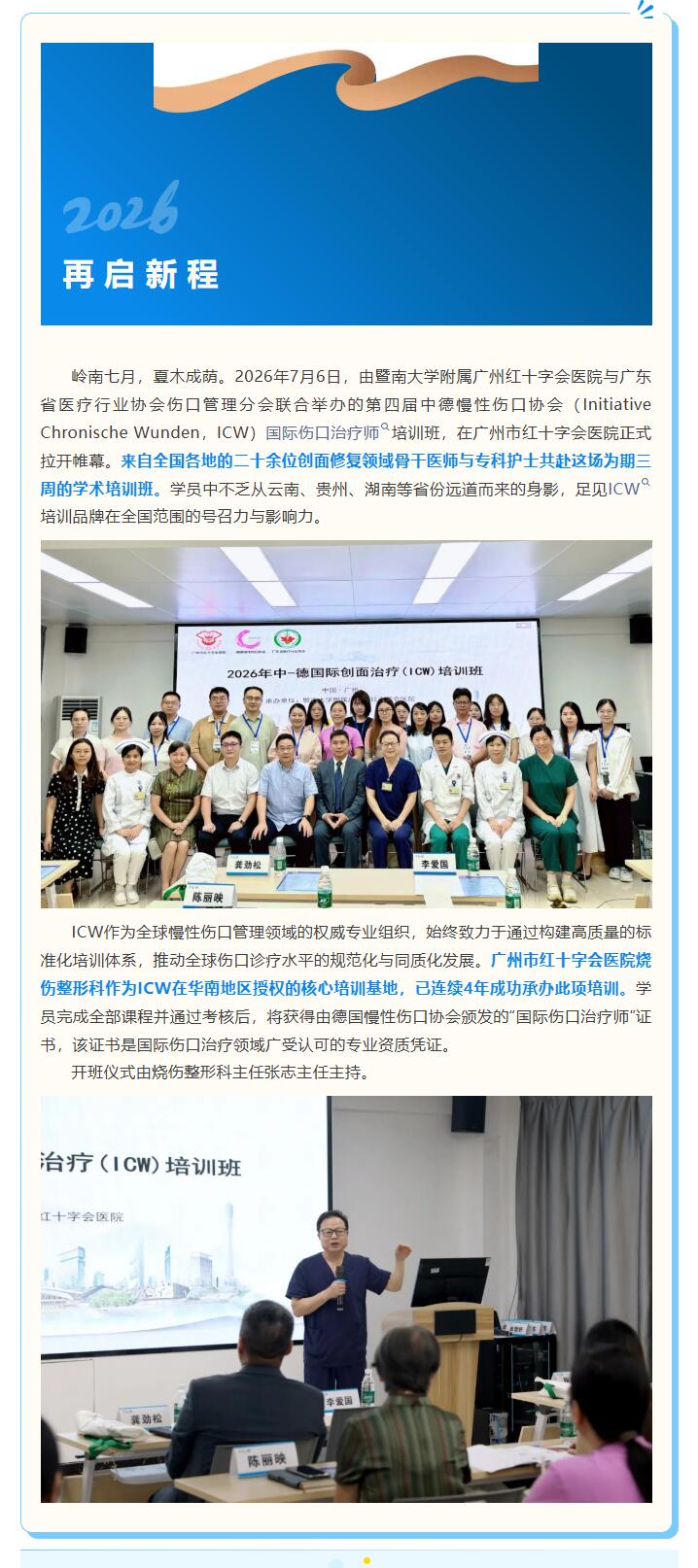
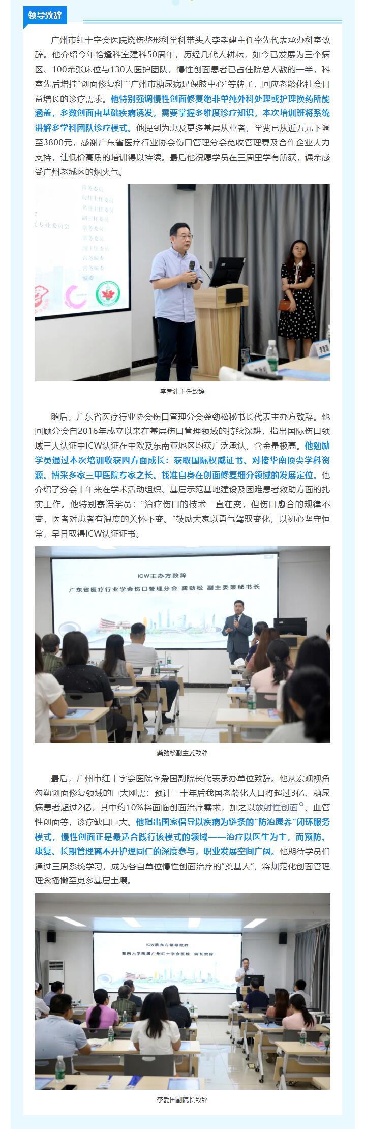
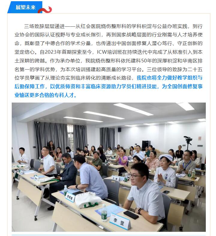
四年深耕,中德携手再谱创面修复新篇章——第四期ICW培训班羊城启幕
本文献转载于红会烧伤,不代表本网站赞同其观点和对其真实性负责,我们只是用于阅读分享,非商业用途,如若侵权,请告知删除。
