版权归中华医学会所有。
未经授权,不得转载、摘编本刊文章。
引用本文:简喜超, 简扬, 邓呈亮. 2025版《中国糖尿病足防治实践指南》解读[J]. 中华医学美学美容杂志, 2026, 32(2): 99-103. DOI: 10.3760/cma.j.cn114657-20251215-00266.
通信作者:邓呈亮,Email:该Email地址已收到反垃圾邮件插件保护。要显示它您需要在浏览器中启用JavaScript。

Athina Stamati1 · Athanasios Christoforidis2
Received: 7 October 2024 / Accepted: 31 December 2024 / Published online: 10 January 2025 © The Author(s) 2025
Abstract
Aims To assess the efficacy and safety of automated insulin delivery (AID) systems compared to standard care in managing glycaemic control during pregnancy in women with Type 1 Diabetes Mellitus (T1DM).
Methods We searched MEDLINE, Cochrane Library, registries and conference abstracts up to June 2024 for randomized controlled trials (RCTs) and observational studies comparing AID to standard care in pregnant women with T1DM. We con-ducted random effects meta-analyses for % of 24-h time in range of 63–140 mg/dL (TIR), time in hyperglycaemia (>140 mg/ dl and>180 mg/dL), hypoglycaemia (<63 mg/dl and<54 mg/dL), total insulin dose (units/kg/day), glycemic variability (%), changes in HbA1c (%), maternal and fetal outcomes.
Results Thirteen studies (450 participants) were included. AID significantly increased TIR (Mean difference, MD 7.01%, 95% CI 3.72–10.30) and reduced time in hyperglycaemia>140 mg/dL and>180 mg/dL (MD – 5.09%, 95% CI – 9.41 to – 0.78 and MD – 2.44%, 95% CI – 4.69 to – 0.20, respectively). Additionally, glycaemic variability was significantly reduced (MD – 1.66%, 95% CI – 2.73 to – 0.58). Other outcomes did not differ significantly.
Conclusion AID systems effectively improve glycaemic control during pregnancy in women with T1DM by increasing TIR and reducing hyperglycaemia without any observed adverse short-term effects on maternal and fetal outcomes.
Keywords Automated insulin delivery · Pregnancy · Type 1 diabetes mellitus · Systematic review · Meta-analysis
Marco Marigliano1,11*† , Roberto Franceschi2†, Enza Mozzillo3†, Valentina Tiberi4†, Monica Marino4 , Giada Boccolini4 , Malgorzata Wasniewska5 , Maria Elizabeth Street6 , Maria Rosaria Licenziati7 , Riccardo Bonfanti8 , Felice Citriniti9 , Giuseppe D’Annunzio10, Maria Carolina Salerno3 , Valentino Cherubini4 and the Diabetes Study Group of the Italian Society of Pediatric Endocrinology and Diabetology (ISPED)
† Marco Marigliano, Roberto Franceschi, Enza Mozzillo and Valentina Tiberi contributed equally to this work.
Backgrounds The incidence of Type 1 Diabetes (T1D) in children and adolescents is increasing by 3–4% per year. Children and adolescents with T1D (CwD) should receive person-centered, specialized treatment from a multidisciplinary team to ensure appropriate care. Italy is the first to implement a countrywide T1D screening program, which will raise the need for funding for specialized pediatric care. The study aims to update the organization of the Italian Centers for pediatric diabetes care.
Methods In 2022, members of the 59 Italian Centers following CwD were invited to complete an email survey regarding the Centers’ organization, characteristics, and activities. The questionnaire included information on responders, department organization, team composition, activities, and the organizational structures: department, ambulatory care services (AC), simple operational units (UOS), simple departmental operational units (UOSd), and complex operational units (UOC).
Results The data collected referred to the year 2022. According to the results, 21,318 people with diabetes were treated. Of these, 19,643 subjects (92.1%) have T1D (16,672 were CwD), 387 (1,8%) have Type 2 Diabetes, and 1,288 (6,1%) have other forms of diabetes. Compared to the 2012 survey, a 13% decrease (from 68 to 59 Centers) in the number of pediatric Centers caring for CwD was observed with a parallel increase of total (+6.6%) and average (+22%) number of CwD per Center. The estimated prevalence of T1D has increased (1.4 vs. 1.7 per 1,000 CwD—2012 vs. 2022). A reduction in numbers for AC (-22%) and UOS (-35%) was observed, whereas UOSd/UOC increased by 50%. Almost 35% of the dietitians and 40% of the psychologists were not permanent members of the multidisciplinary diabetes team.
Conclusions The observed decrease in the overall number of pediatric diabetes Centers, the reduction in specialized and dedicated HCPs, and the concurrent increase in the number of treated CwD in the last ten years indicate an alarming situation for pediatric diabetes treatment in Italy. Furthermore, the projected rise in CwD due to the National T1D screening program emphasizes the need for increased resources for specialized pediatric care of CwD at all stages.
Keywords Type 1 diabetes, Care, Children, Adolescents, Benchmarking, Team, Screening, Technology
Loϊc Binan,1,2 Aiping Jiang,3,4,5 Serwah A. Danquah,1,6,12 Vera Valakh,1,6,13 Brooke Simonton,2,14 Jon Bezney,2,15 Robert T. Manguso,3,4,5 Kathleen B. Yates,3,4,5 Ralda Nehme,6 Brian Cleary,7,8,9,10,11,* and Samouil L. Farhi1,16,*
1 Spatial Technology Platform, Broad Institute of MIT and Harvard, Cambridge, MA 02142, USA
2 Klarman Cell Observatory, Broad Institute of MIT and Harvard, Cambridge, MA 02142, USA
3 Cancer Program, Broad Institute of Harvard and MIT, Cambridge, MA 02142, USA
4 Krantz Family Center for Cancer Research, Massachusetts General Hospital, Boston, MA 02144, USA
5 Department of Medicine, Harvard Medical School, Boston, MA 02115, USA
6 Stanley Center for Psychiatric Research, Broad Institute of MIT and Harvard, Cambridge, MA 02142, USA
7 Faculty of Computing and Data Sciences, Boston University, Boston, MA 02215, USA
8 Department of Biology, Boston University, Boston, MA 02215, USA
9 Department of Biomedical Engineering, Boston University, Boston, MA 02215, USA
10 Program in Bioinformatics, Boston University, Boston, MA 02215, USA
11 Biological Design Center, Boston University, Boston, MA 02215, USA
12 Present address: Department of Laboratory Medicine and Pathology, University of Washington, Seattle, WA, USA
13 Present address: Stoke Therapeutics, Bedford, MA, USA
14 Present address: The Ken & Ruth Davee Department of Neurology, Northwestern University Feinberg School of Medicine, Chicago, IL, USA
15 Present address: Department of Genetics, Stanford University School of Medicine, Stanford, CA, USA
16 Lead contact
*Correspondence: 该Email地址已收到反垃圾邮件插件保护。要显示它您需要在浏览器中启用JavaScript。 (B.C.), 该Email地址已收到反垃圾邮件插件保护。要显示它您需要在浏览器中启用JavaScript。 (S.L.F.)
https://doi.org/10.1016/j.cell.2025.02.012
SUMMARY
Pooled optical screens have enabled the study of cellular interactions, morphology, or dynamics at massive scale, but they have not yet leveraged the power of highly plexed single-cell resolved transcriptomic readouts to inform molecular pathways. Here, we present a combination of imaging spatial transcriptomics with parallel optical detection of in situ amplified guide RNAs (Perturb-FISH). Perturb-FISH recovers intracellular effects that are consistent with single-cell RNA-sequencing-based readouts of perturbation effects (Perturb-seq) in a screen of lipopolysaccharide response in cultured monocytes, and it uncovers intercellular and density-dependent regulation of the innate immune response. Similarly, in three-dimensional xenograft models, Perturb-FISH identifies tumor-immune interactions altered by genetic knockout. When paired with a functional readout in a separate screen of autism spectrum disorder risk genes in human-induced pluripotent stem cell (hIPSC) astrocytes, Perturb-FISH shows common calcium activity phenotypes and their associated genetic interactions and dysregulated molecular pathways. Perturb-FISH is thus a general method for studying the genetic and molecular associations of spatial and functional biology at single-cell resolution.
Xiaoqian Liu,1,5,6,7,21 Zunpeng Liu,1,5,7,21 Zeming Wu,2,5,7,21 Jie Ren,3,5,7,21 Yanling Fan,3,7 Liang Sun,9 Gang Cao,8 Yuyu Niu,11,12,13 Baohu Zhang,1,7 Qianzhao Ji,2,7 Xiaoyu Jiang,2,7 Cui Wang,3,7 Qiaoran Wang,3,7 Zhejun Ji,1,5,7 Lanzhu Li,2,7 Concepcion Rodriguez Esteban,18 Kaowen Yan,2,5,7 Wei Li,4 Yusheng Cai,2,5,7 Si Wang,4,5,7,15,16 Aihua Zheng,7,19 Yong E. Zhang,7,14 Shengjun Tan,14 Yingao Cai,7,14 Moshi Song,2,5,6,7 Falong Lu,7,10 Fuchou Tang,17 Weizhi Ji,11,12,20 Qi Zhou,1,5,6,7,20 Juan Carlos Izpisua Belmonte,18,20 Weiqi Zhang,3,5,7,* Jing Qu,1,5,6,7,* and Guang-Hui Liu2,4,5,6,7,22,*
1State Key Laboratory of Stem Cell and Reproductive Biology, Institute of Zoology, Chinese Academy of Sciences, Beijing 100101, China
2 State Key Laboratory of Membrane Biology, Institute of Zoology, Chinese Academy of Sciences, Beijing 100101, China
3 CAS Key Laboratory of Genomic and Precision Medicine, Beijing Institute of Genomics, Chinese Academy of Sciences and China National Center for Bioinformation, Beijing 100101, China
4 Advanced Innovation Center for Human Brain Protection, National Clinical Research Center for Geriatric Disorders, Xuanwu Hospital Capital Medical University, Beijing 100053, China
5 Institute for Stem cell and Regeneration, Chinese Academy of Sciences, Beijing 100101, China
6 Beijing Institute for Stem Cell and Regenerative Medicine, Beijing 100101, China
7 University of Chinese Academy of Sciences, Beijing 100049, China
8 State Key Laboratory of Agricultural Microbiology, Huazhong Agricultural University, Wuhan 430070, China
9 NHC Beijing Institute of Geriatrics, NHC Key Laboratory of Geriatrics, Institute of Geriatric Medicine of Chinese Academy of Medical Sciences, National Center of Gerontology/Beijing Hospital, Beijing 100730, China
10 State Key Laboratory of Molecular Developmental Biology, Institute of Genetics and Developmental Biology, The Innovative Academy of Seed Design, Chinese Academy of Sciences, Beijing 100101, China
11 State Key Laboratory of Primate Biomedical Research, Institute of Primate Translational Medicine, Kunming University of Science and Technology, Kunming, Yunnan 650500, China
12 Yunnan Key Laboratory of Primate Biomedical Research, Kunming, Yunnan 650500, China
13 Faculty of Life Science and Technology, Kunming University of Science and Technology, Kunming, Yunnan 650500, China
14 Key Laboratory of Zoological Systematics and Evolution, Institute of Zoology, Chinese Academy of Sciences, Beijing 100101, China
15 Aging Translational Medicine Center, International Center for Aging and Cancer, Xuanwu Hospital, Capital Medical University, Beijing Municipal Geriatric Medical Research Center, Beijing 100053, China
16 The Fifth People’s Hospital of Chongqing, Chongqing 400062, China
17 Peking-Tsinghua Center for Life Sciences, Peking University, Beijing 100871, China
18 Altos Labs, Inc., San Diego, CA 94022, USA
19 State Key Laboratory of Integrated Management of Pest Insects and Rodents, Institute of Zoology, Chinese Academy of Sciences, Beijing 100101, China
20 Senior author
21 These authors contributed equally
22 Lead contact
*Correspondence: 该Email地址已收到反垃圾邮件插件保护。要显示它您需要在浏览器中启用JavaScript。 (W.Z.), 该Email地址已收到反垃圾邮件插件保护。要显示它您需要在浏览器中启用JavaScript。 (J.Q.), 该Email地址已收到反垃圾邮件插件保护。要显示它您需要在浏览器中启用JavaScript。 (G.-H.L.)
https://doi.org/10.1016/j.cell.2022.12.017
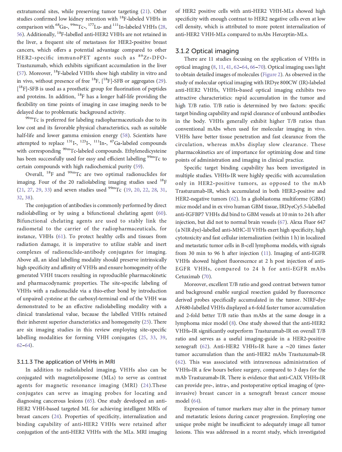
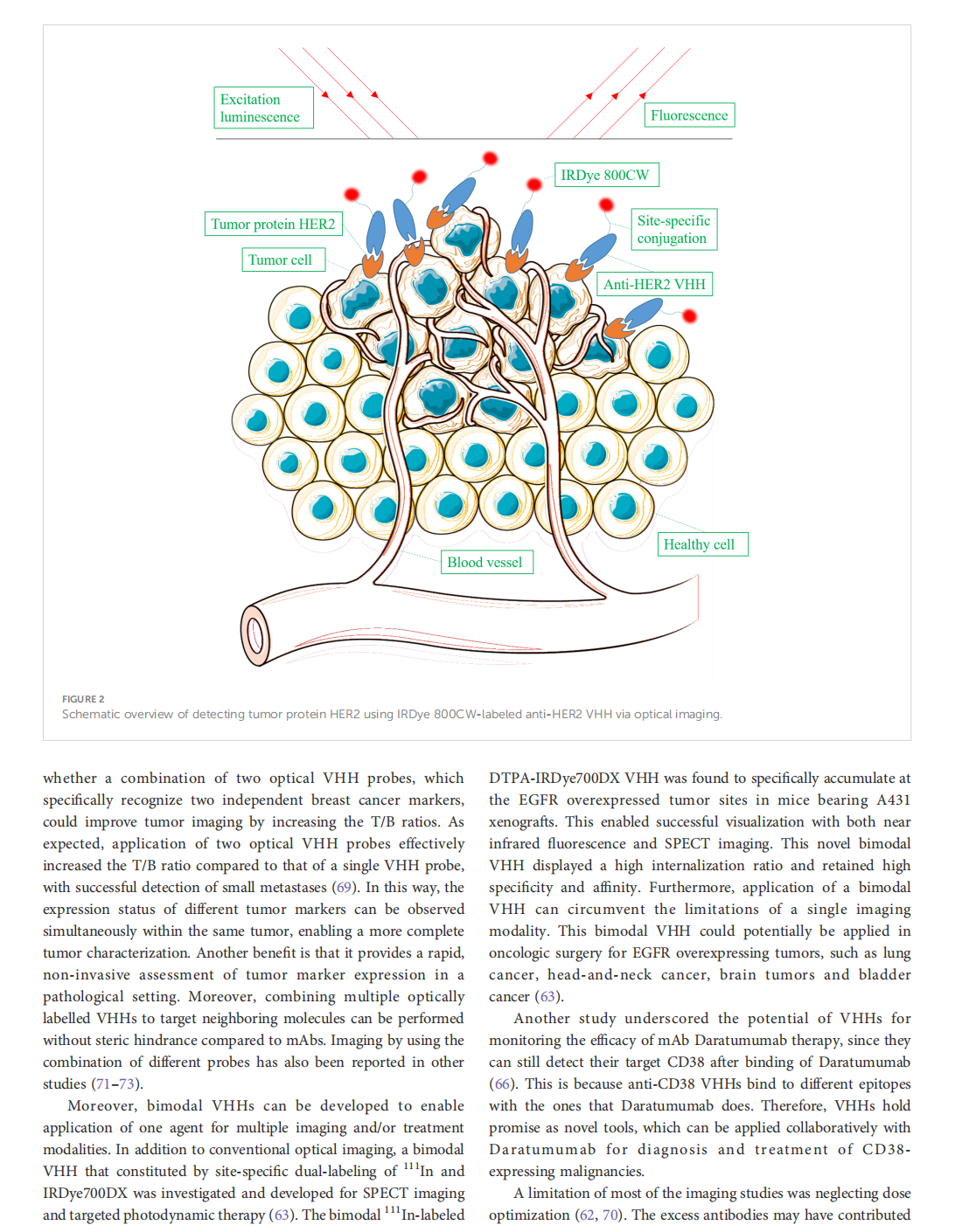
Whether and how certain transposable elements with viral origins, such as endogenous retroviruses (ERVs) dormant in our genomes, can become awakened and contribute to the aging process is largely unknown. In human senescent cells, we found that HERVK (HML-2), the most recently integrated human ERVs, are unlocked to transcribe viral genes and produce retrovirus-like particles (RVLPs). These HERVK RVLPs constitute a transmissible message to elicit senescence phenotypes in young cells, which can be blocked by neutralizing antibodies. The activation of ERVs was also observed in organs of aged primates and mice as well as in human tissues and serum from the elderly. Their repression alleviates cellular senescence and tissue degeneration and, to some extent, organismal aging. These findings indicate that the resurrection of ERVs is a hallmark and driving force of cellular senescence and tissue aging.
Mateo Ve´ lez-Fort,1,3 Lee Cossell,1,3 Laura Porta,1 Claudia Clopath,1,2 and Troy W. Margrie1,4,*
1 Sainsbury Wellcome Centre, University College London, London, UK
2 Bioengineering Department, Imperial College London, London, UK
3 These authors contributed equally
4 Lead contact
*Correspondence: 该Email地址已收到反垃圾邮件插件保护。要显示它您需要在浏览器中启用JavaScript。
https://doi.org/10.1016/j.cell.2025.01.032
Knowing whether we are moving or something in the world is moving around us is possibly the most critical sensory discrimination we need to perform. How the brain and, in particular, the visual system solves this motion-source separation problem is not known. Here, we find that motor, vestibular, and visual motion signals are used by the mouse primary visual cortex (VISp) to differentially represent the same visual flow information according to whether the head is stationary or experiencing passive versus active translation. During locomotion, we find that running suppresses running-congruent translation input and that translation signals dominate VISp activity when running and translation speed become incongruent. This cross-modal interaction between the motor and vestibular systems was found throughout the cortex, indicating that running and translation signals provide a brain-wide egocentric reference frame for computing the internally generated and actual speed of self when moving through and sensing the external world.
Ugne Klibaite,1,4,* Tianqing Li,2,4 Diego Aldarondo,1,3 Jumana F. Akoad,1 Bence P. O¨ lveczky,1,* and Timothy W. Dunn2,5,*
1 Department of Organismic and Evolutionary Biology, Harvard University, Cambridge, MA 02138, USA
2 Department of Biomedical Engineering, Duke University, Durham, NC 27708, USA
3 Present address: Fauna Robotics, New York, NY 10003, USA
4 These authors contributed equally
5 Lead contact
*Correspondence: 该Email地址已收到反垃圾邮件插件保护。要显示它您需要在浏览器中启用JavaScript。 (U.K.), 该Email地址已收到反垃圾邮件插件保护。要显示它您需要在浏览器中启用JavaScript。 (B.P.O¨ .), 该Email地址已收到反垃圾邮件插件保护。要显示它您需要在浏览器中启用JavaScript。 (T.W.D.)
https://doi.org/10.1016/j.cell.2025.01.044
Social interaction is integral to animal behavior. However, lacking tools to describe it in quantitative and rigorous ways has limited our understanding of its structure, underlying principles, and the neuropsychiatric disorders, like autism, that perturb it. Here, we present a technique for high-resolution 3D tracking of postural dynamics and social touch in freely interacting animals, solving the challenging subject occlusion and partassignment problems using 3D geometric reasoning, graph neural networks, and semi-supervised learning. We collected over 110 million 3D pose samples in interacting rats and mice, including seven monogenic autism rat lines. Using a multi-scale embedding approach, we identified a rich landscape of stereotyped actions, interactions, synchrony, and body contacts. This high-resolution phenotyping revealed a spectrum of changes in autism models and in response to amphetamine not resolved by conventional measurements. Our framework and large library of interactions will facilitate studies of social behaviors and their neurobiological underpinnings.
伤口世界平台生态圈,以“关爱人间所有伤口患者”为愿景,连接、整合和拓展线上和线下的管理慢性伤口的资源,倡导远程、就近和居家管理慢性伤口,解决伤口专家的碎片化时间的价值创造、诊疗经验的裂变复制、和患者的就近、居家和低成本管理慢性伤口的问题。
2019广东省医疗行业协会伤口管理分会年会
扫一扫了解详情:
任何关于疾病的建议都不能替代执业医师的面对面诊断。所有门诊时间仅供参考,最终以医院当日公布为准。
网友、医生言论仅代表其个人观点,不代表本站同意其说法,请谨慎参阅,本站不承担由此引起的法律责任。