
This article is excerpted from the Histochemistry and Cell Biology (2024) 162:259–271 by Wound World.

Marta Arnal‑Forné1 · Tamara Molina‑García2 · María Ortega2 · Víctor Marcos‑Garcés2,3 · Pilar Molina4 · Antonio Ferrández‑Izquierdo1,2,5 · Pilar Sepulveda1,6,7 · Vicente Bodí2,3,6,8 · César Ríos‑Navarro1,2,6 · Amparo Ruiz‑Saurí1,2,6
Accepted: 10 June 2024 / Published online: 2 July 2024
© The Author(s) 2024
Abstract
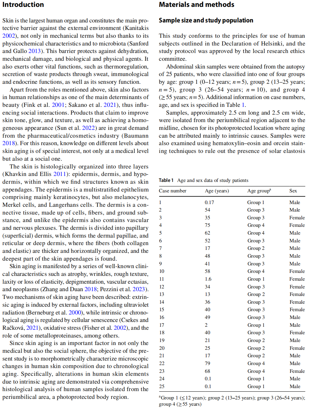
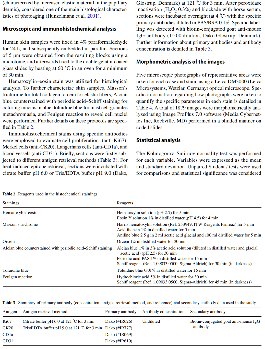
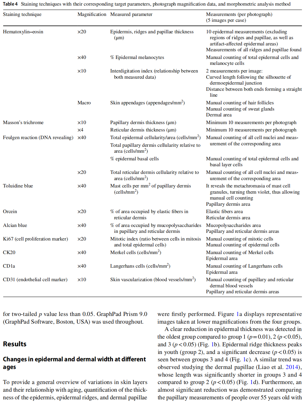
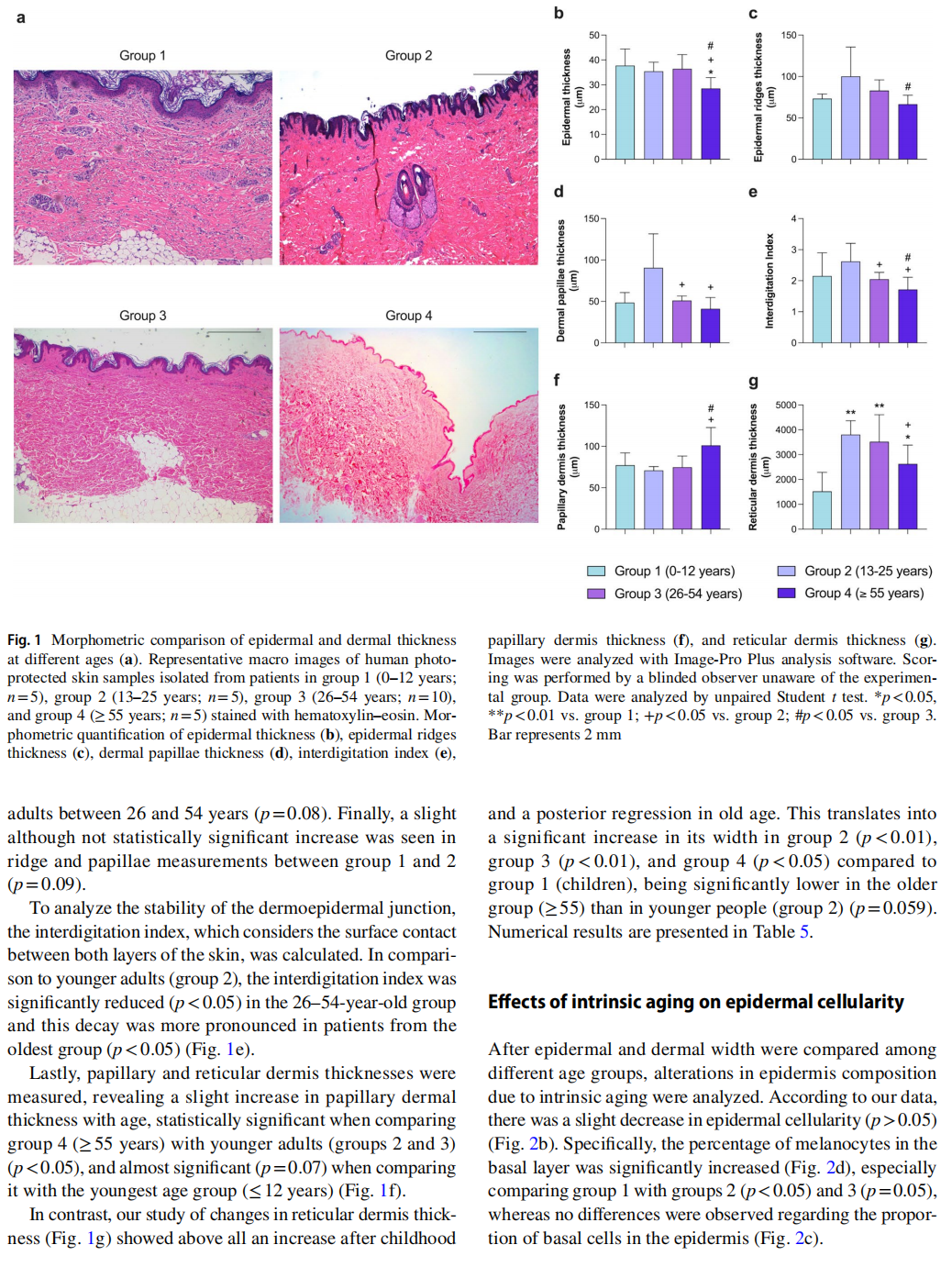
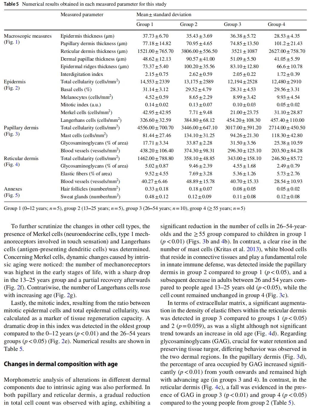
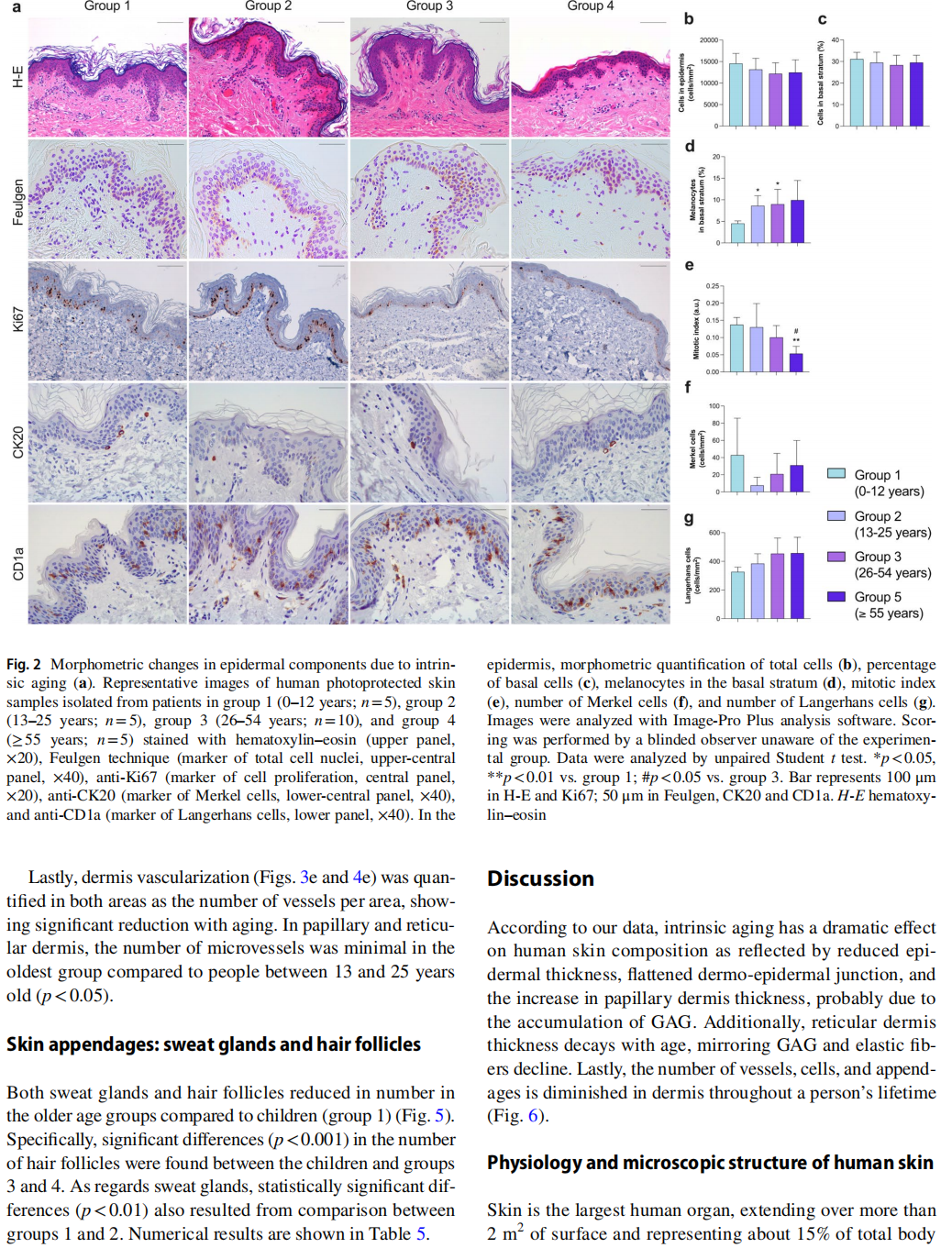
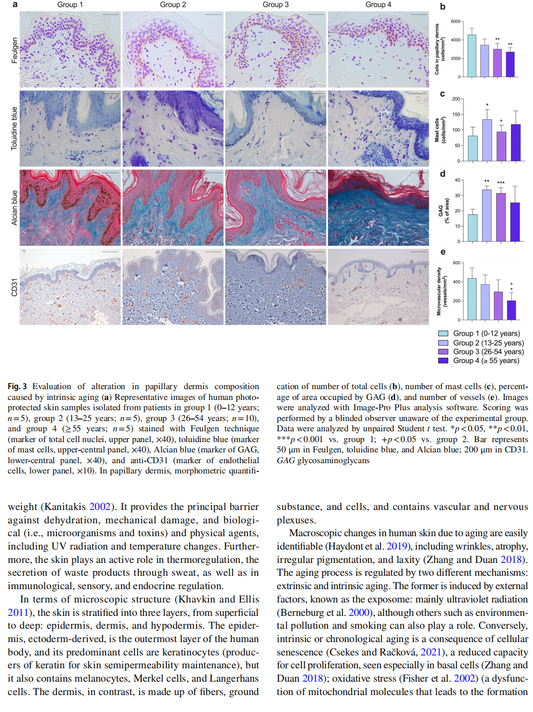
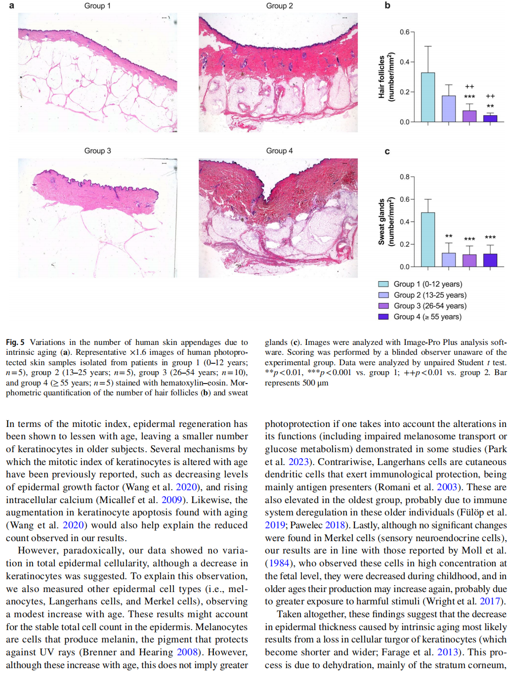
Skin represents the main barrier against the external environment, but also plays a role in human relations, as one of the prime determinants of beauty, resulting in a high consumer demand for skincare-related pharmaceutical products. Given the importance of skin aging in both medical and social spheres, the present research aims to characterize microscopic changes in human skin composition due to intrinsic aging (as opposed to aging infuenced by external factors) via histological analysis of a photoprotected body region. Samples from 25 autopsies were taken from the periumbilical area and classifed into four age groups: group 1 (0–12 years), group 2 (13–25 years), group 3 (26–54 years), and group 4 (≥55 years). Diferent traditional histological (hematoxylin–eosin, Masson’s trichrome, orcein, toluidine, Alcian blue, and Feulgen reaction) and immunohistochemical (CK20, CD1a, Ki67, and CD31) stains were performed. A total of 1879 images photographed with a Leica DM3000 optical microscope were morphometrically analyzed using Image ProPlus 7.0 for further statistical analysis with GraphPad 9.0. Our results showed a reduction in epidermis thickness, interdigitation and mitotic indexes, while melanocyte count was raised. Papillary but not reticular dermis showed increased thickness with aging. Specifcally, in the papillary layer mast cells and glycosaminoglycans were expanded, whereas the reticular dermis displayed a diminution in glycosaminoglycans and elastic fbers. Moreover, total cellularity and vascularization of both dermises were diminished with aging. This morphometric analysis of photoprotected areas reveals that intrinsic aging signifcantly infuences human skin composition. This study paves the way for further research into the molecular basis underpinning these alterations, and into potential antiaging strategies.
Keywords Skin aging · Intrinsic aging · Morphometric analysis · Human biopsies
César Ríos-Navarro
该Email地址已收到反垃圾邮件插件保护。要显示它您需要在浏览器中启用JavaScript。
* Amparo Ruiz-Saurí 该Email地址已收到反垃圾邮件插件保护。要显示它您需要在浏览器中启用JavaScript。; 该Email地址已收到反垃圾邮件插件保护。要显示它您需要在浏览器中启用JavaScript。
1 Department of Pathology, University of Valencia, Avda. Blasco Ibáñez 15. 46010, Valencia, Spain
2 Instituto de Investigación Sanitaria INCLIVA Biomedical Research Institute, Avda. Menéndez Pelayo 4acc,
46010 Valencia, Spain
3 Cardiology Department, Hospital Clínico Universitario, Valencia, Spain
4 Department of Pathology, Instituto de Medicina Legal y Ciencias Forenses, Valencia, Spain
5 Anatomic Pathology Department, Hospital Clínico Universitario, Valencia, Spain
6 Centro de Investigación Biomédica en Red (CIBER)-CV, Madrid, Spain
7 Regenerative Medicine and Heart Transplantation Unit, Instituto de Investigación Sanitaria La Fe, Valencia, Spain
8 Department of Medicine, University of Valencia, Valencia, Spain
This article is excerpted from the Histochemistry and Cell Biology (2024) 162:259–271 by Wound World.