
伤口世界
电子邮件地址: 该Email地址已收到反垃圾邮件插件保护。要显示它您需要在浏览器中启用JavaScript。
- 星期四, 15 1月 2026
Bridging clinic to home: domestic devices in dermatological diagnostics and treatments
This article is excerpted from the 《Frontiers in Digital Health》 by Wound World
- 星期三, 14 1月 2026
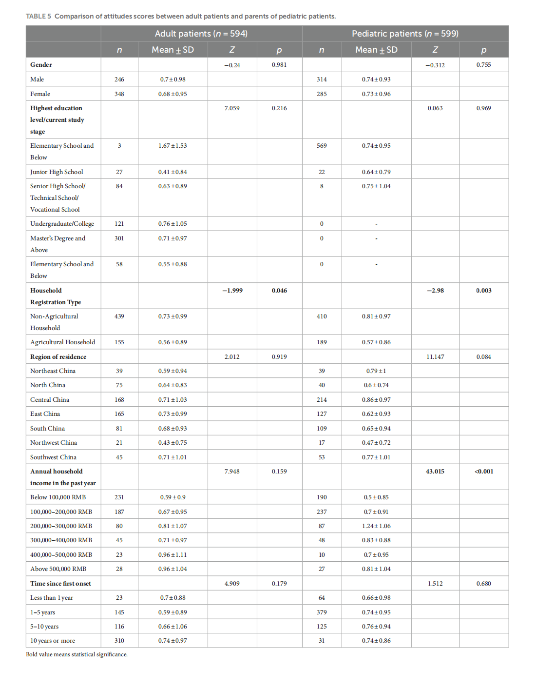
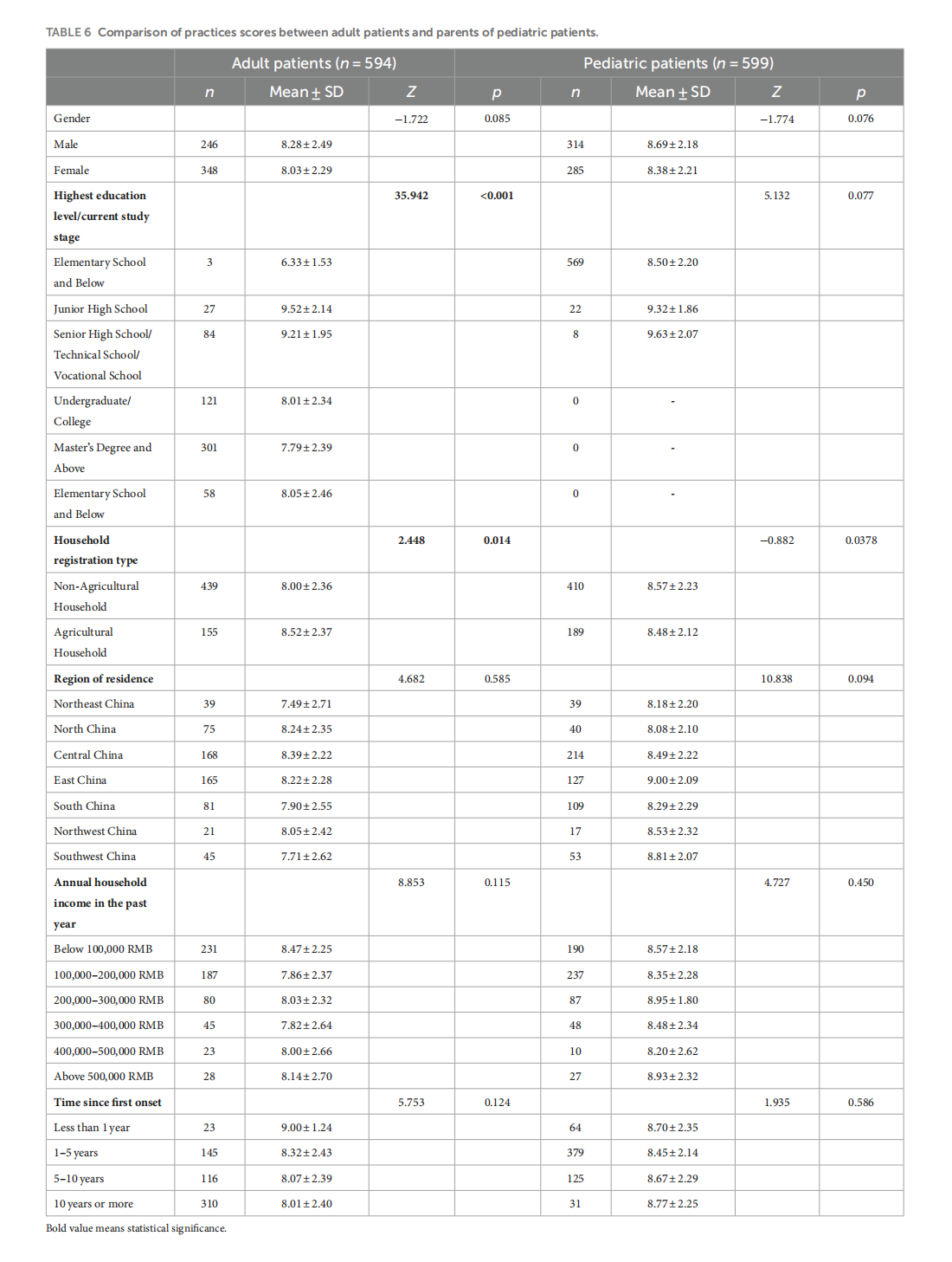
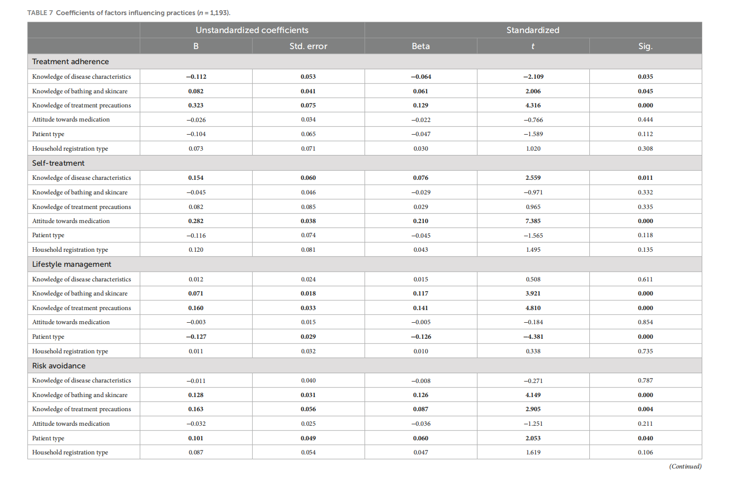
Knowledge, attitudes, and practices in adult patients and parents of pediatric atopic dermatitis patients: a cross-sectional study
This article is excerpted from the《Frontiers in Public Health》 by Wound World

- 星期二, 13 1月 2026
Spectrum of sensitive skin in India: a collaborative expert position statement
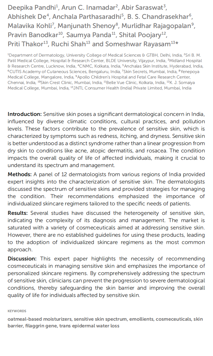
This article is excerpted from the 《Frontiers in Medicine》 by Wound World
- 星期一, 12 1月 2026
Medical Cannabis for the Treatment of Migraine in Adults: A Review of the Evidence
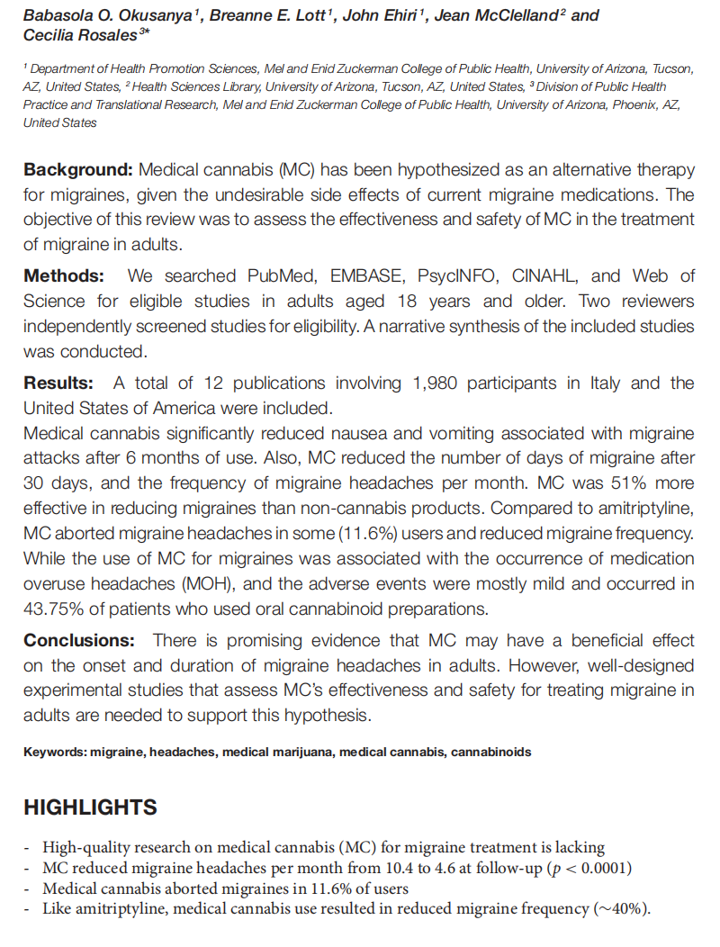
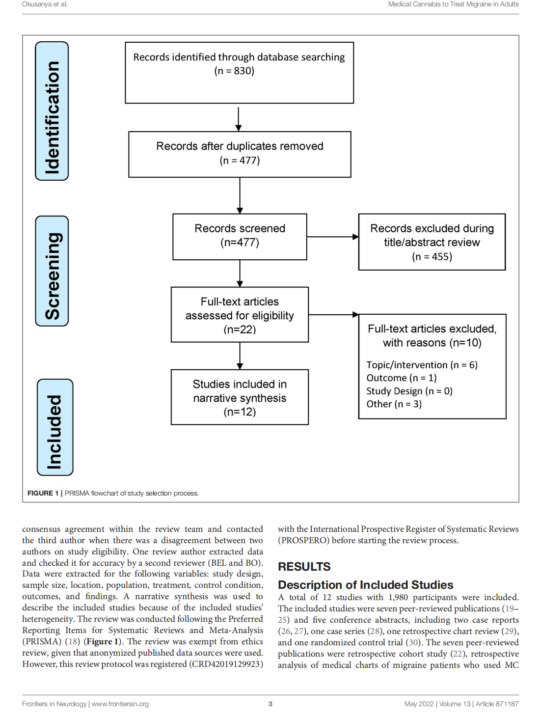
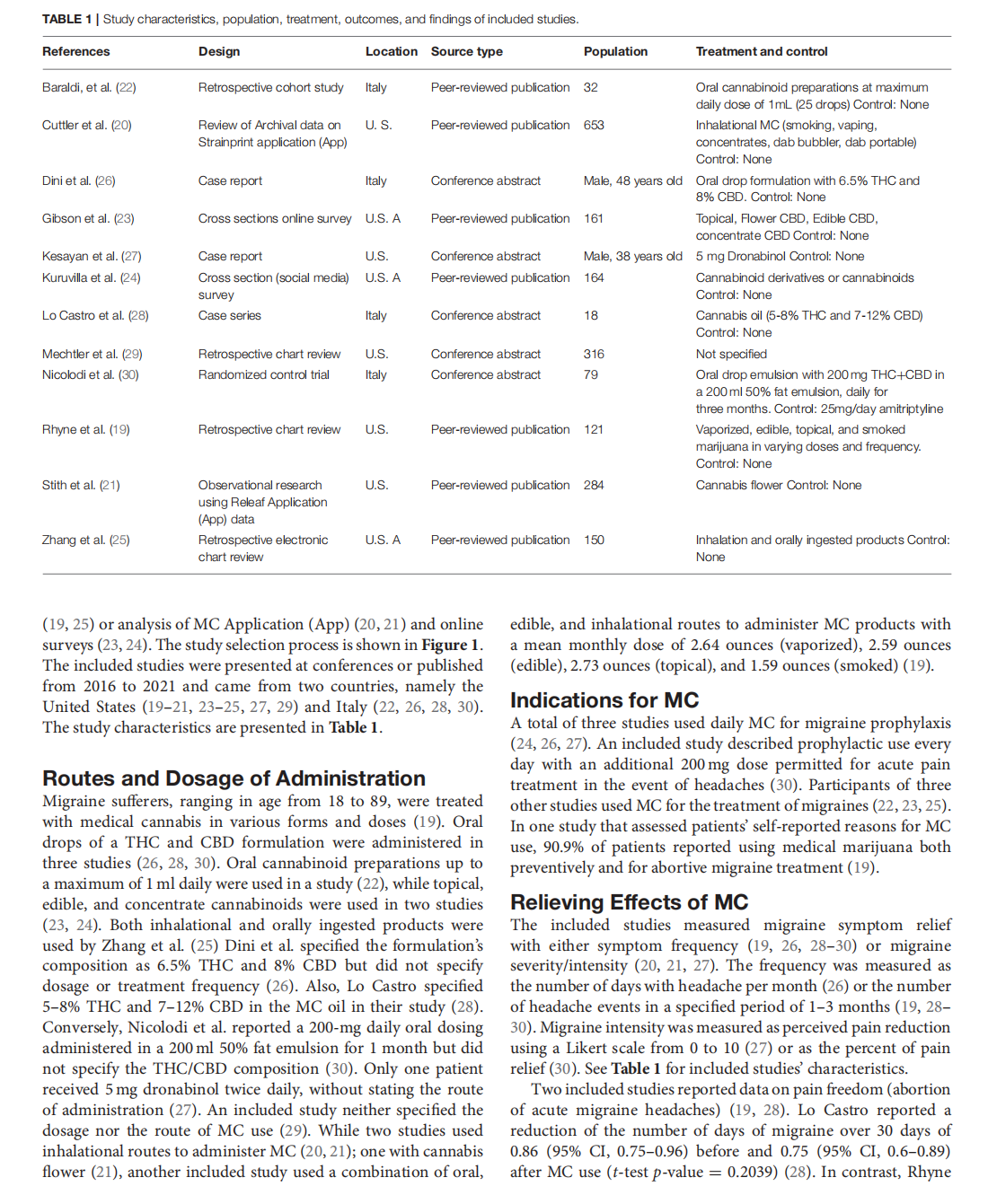
This article is excerpted from the《Frontiers in Neurology》by Wound World
- 星期五, 09 1月 2026
Integrative population pharmacokinetic/ pharmacodynamic analysis of nemonoxacin capsule in Chinese patients with community-acquired pneumonia
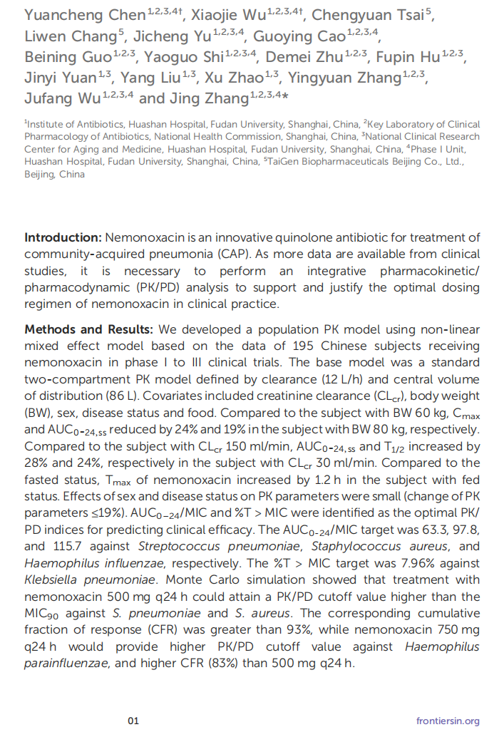
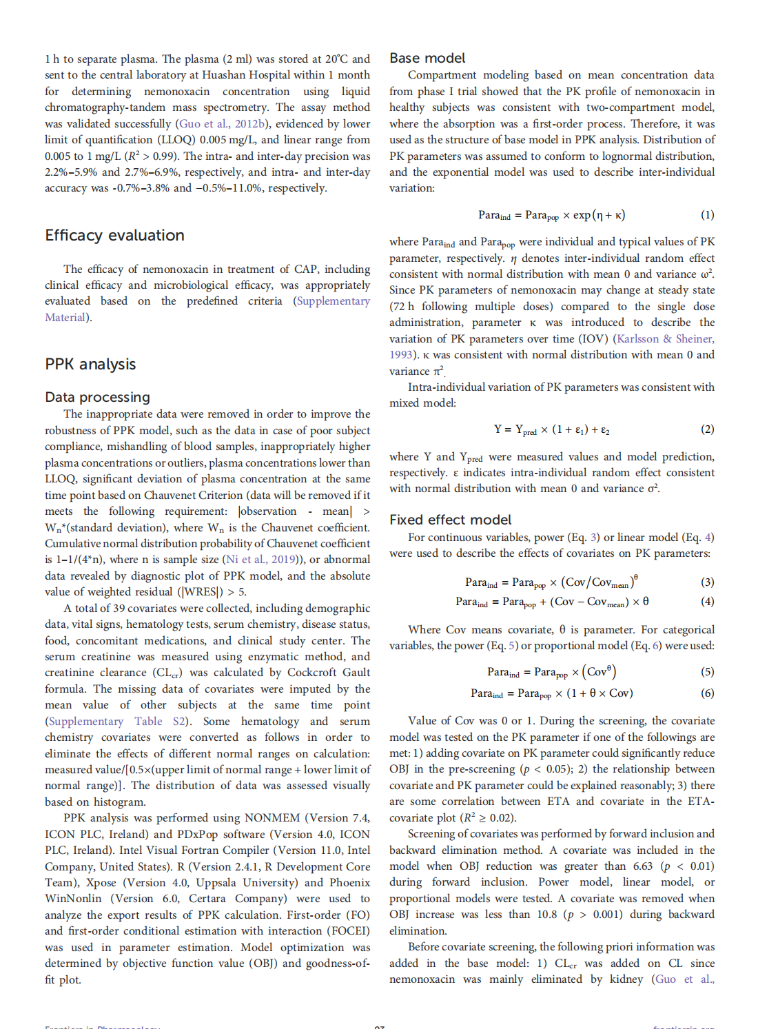
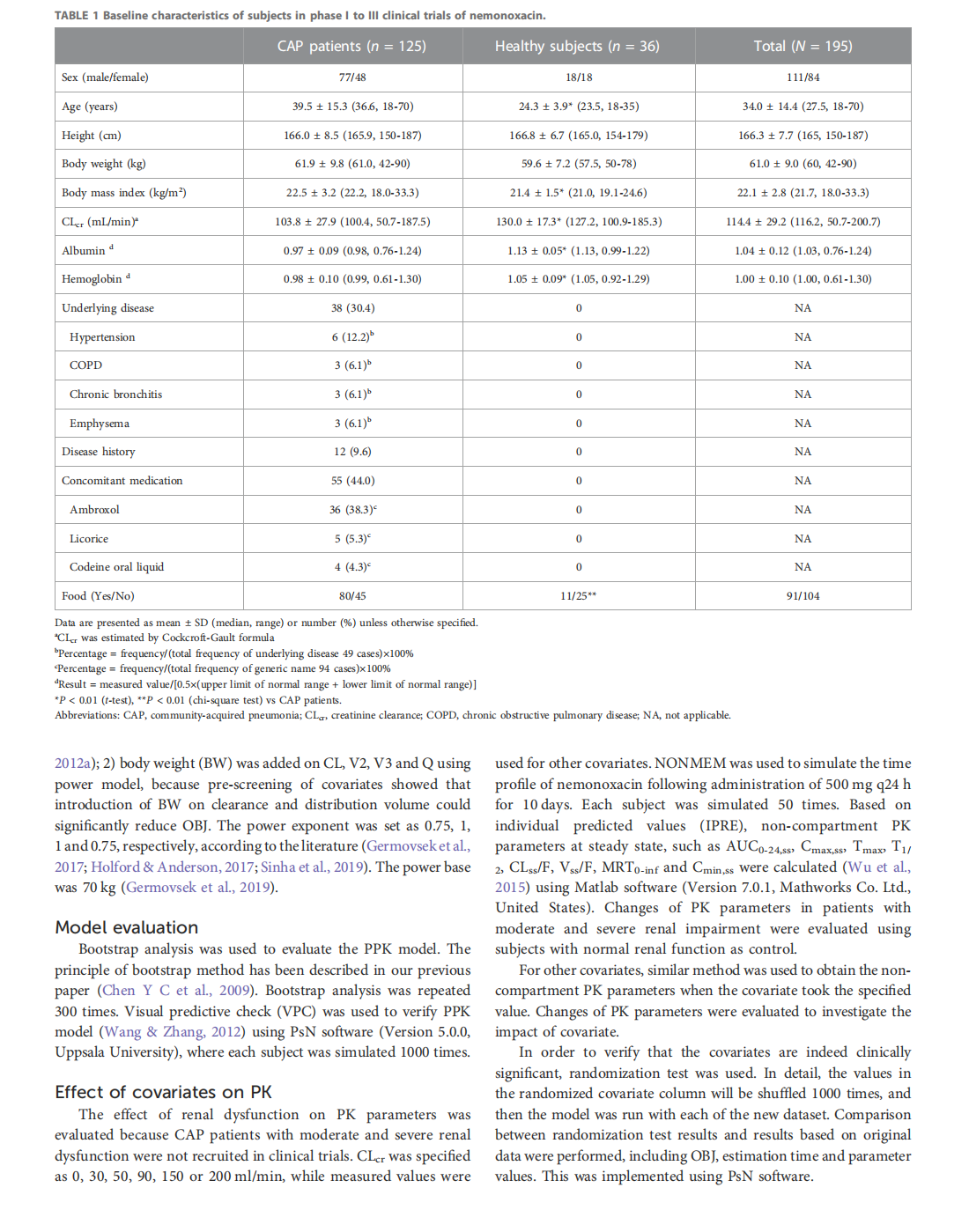
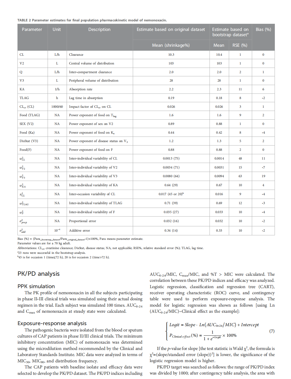
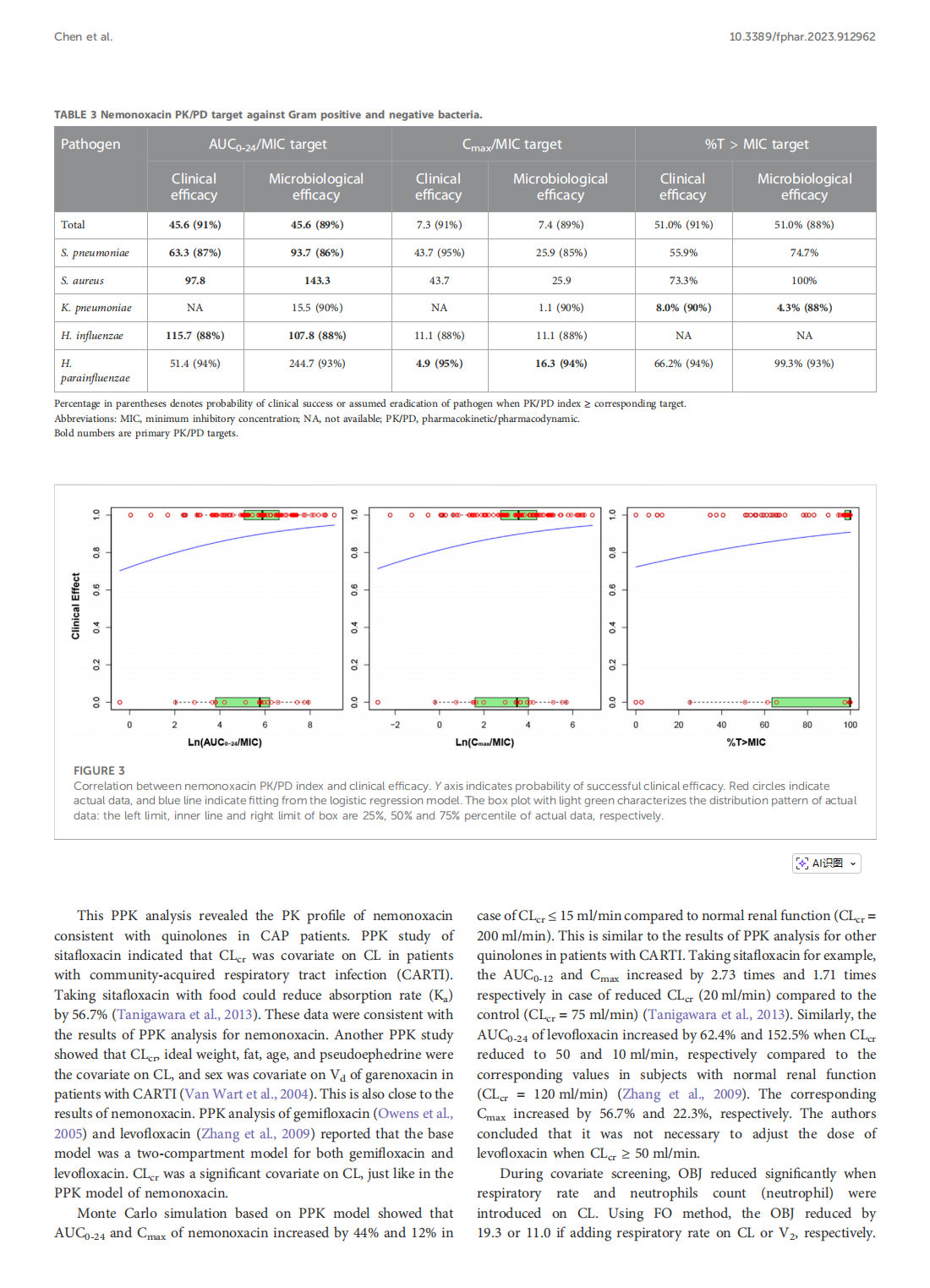
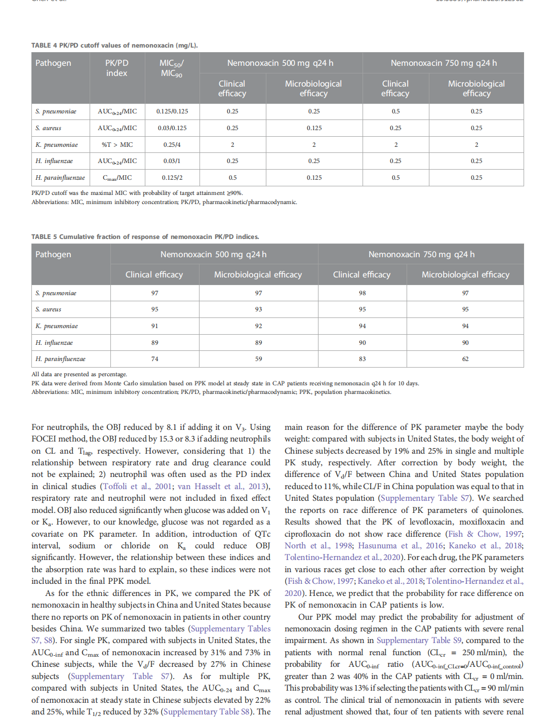
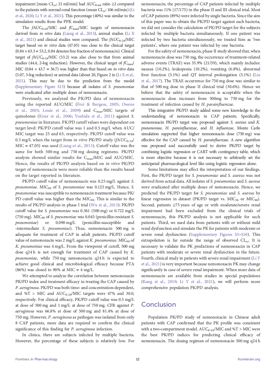
This article is excerpted from the《Frontiers in Pharmacology》by Wound World
- 星期四, 08 1月 2026
Construction of a management and prevention program for targeted therapy-induced hand-foot skin reaction
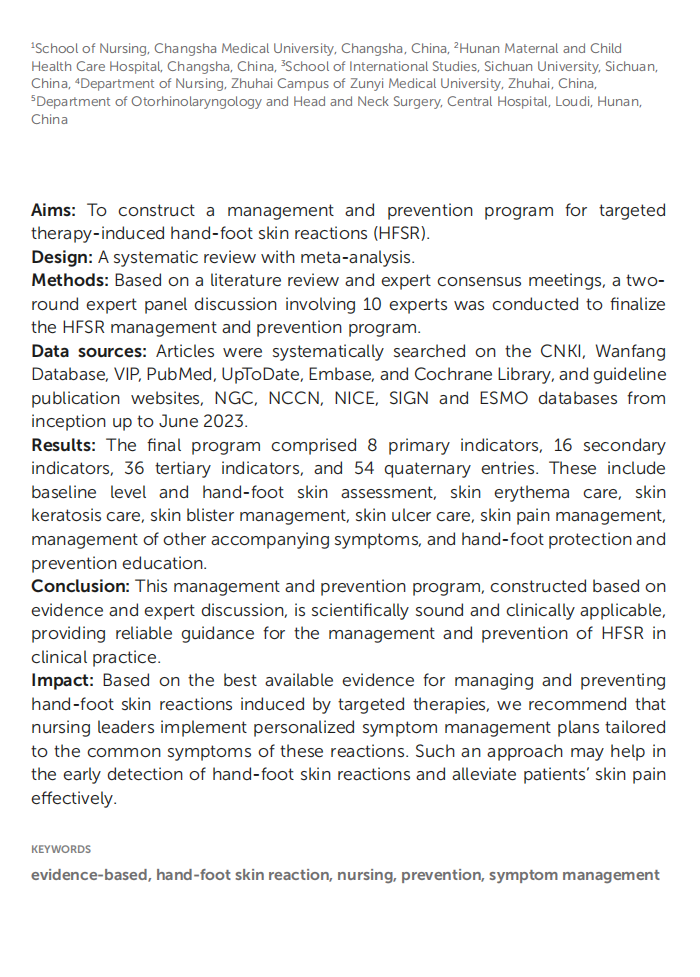


This article is excerpted from the《Frontiers in Oncology》by Wound World
