
伤口世界

- 星期一, 02 9月 2024
LINE1 Derepression in Aged Wild-Type and SIRT6- Deficient Mice Drives Inflammation
Authors
Matthew Simon, Michael Van Meter, Julia Ablaeva, ..., John M. Sedivy, Andrei Seluanov, Vera Gorbunova
Correspondence
该Email地址已收到反垃圾邮件插件保护。要显示它您需要在浏览器中启用JavaScript。 (A.S.), 该Email地址已收到反垃圾邮件插件保护。要显示它您需要在浏览器中启用JavaScript。 (V.G.)
In Brief
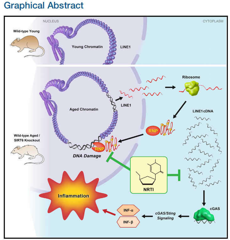
Simon et al. show that LINE1 retrotransposon elements are derepressed in aged and progeroid mice. Cytoplasmic accumulation of LINE1 cDNA copies induced a type I interferon response, through the cGAS DNA sensing pathway, resulting in pathological inflammation. Inhibiting L1 replication significantly improved the health and lifespan of aged mice.
Highlights
● SIRT6 KO mice accumulate L1 cDNA, triggering interferon response via cGAS pathway
● Wild-type aged mice accumulate L1 cDNA and display type I interferon response
● Reverse-transcriptase inhibitors rescue type I interferon response and DNA damage
● Reverse-transcriptase inhibitors extend lifespan and improve health of SIRT6 KO mice
SUMMARY
Mice deficient for SIRT6 exhibit a severely shortened lifespan, growth retardation, and highly elevated LINE1 (L1) activity. Here we report that SIRT6-deficient cells and tissues accumulate abundant cytoplasmic L1 cDNA, which triggers strong type I interferon response via activation of cGAS. Remarkably, nucleoside reverse-transcriptase inhibitors (NRTIs), which inhibit L1 retrotransposition, significantly improved health and lifespan of SIRT6 knockout mice and completely rescued type I interferon response. In tissue culture, inhibition of L1 with siRNA or NRTIs abrogated type I interferon response, in addition to a significant reduction of DNA damage markers. These results indicate that L1 activation contributes to the pathologies of SIRT6 knockout mice. Similarly, L1 transcription, cytoplasmic cDNA copy number, and type I interferons were elevated in the wild-type aged mice. As sterile inflammation is a hallmark of aging, we propose that modulating L1 activity may be an important strategy for attenuating age-related pathologies.
Context and Significance
Mammalian aging is complex and likely reflects accumulated damage to our genes/DNA. Retrotransposons are a special class of parasitic genetic elements that can replicate their DNA within our genes, at times amounting to up to 20% of human DNA. Retrotransposons, such as the commonly occurring L1, have been associated with aging, neurodegeneration, and cancer. University of Rochester scientists uncovered L1 retrotransposons as the culprit in many aspects of accelerated aging in mice, a model for human aging. They also linked these special gene elements to inflammation. Experimentally blocking retrotransposon amplification improved the health and lifespan of mice. Although there is a long road ahead, inhibiting retrotransposon activity, and the related inflammation, could eventually be a therapy for age-related diseases.

- 星期五, 30 8月 2024
Cytoplasmic DNA: sources, sensing, and role in aging and disease
Karl N. Miller,1,6 Stella G. Victorelli,2,3,6 Hanna Salmonowicz,2,3,4,5 Nirmalya Dasgupta,1 Tianhui Liu,1 Joa˜o F. Passos,2,3,* and Peter D. Adams1,* 1Aging, Cancer and Immuno-oncology Program, Sanford Burnham Prebys Medical Discovery Institute, La Jolla, CA 92037, USA 2Department of Physiology and Biomedical Engineering, Mayo Clinic, Rochester, MN 55905, USA 3Robert and Arlene Kogod Center on Aging, Mayo Clinic, Rochester, MN 55905, USA 4Institute for Cell and Molecular Biosciences & Newcastle University Institute for Ageing, Newcastle upon Tyne NE4 5PL, UK 5International Institute of Molecular Mechanisms and Machines, Polish Academy of Sciences, 02-109 Warsaw, Poland 6These authors contributed equally
*Correspondence: 该Email地址已收到反垃圾邮件插件保护。要显示它您需要在浏览器中启用JavaScript。 (J.F.P.), 该Email地址已收到反垃圾邮件插件保护。要显示它您需要在浏览器中启用JavaScript。 (P.D.A.)
https://doi.org/10.1016/j.cell.2021.09.034
SUMMARY
Endogenous cytoplasmic DNA (cytoDNA) species are emerging as key mediators of inflammation in diverse physiological and pathological contexts. Although the role of endogenous cytoDNA in innate immune activation is well established, the cytoDNA species themselves are often poorly characterized and difficult to distinguish, and their mechanisms of formation, scope of function and contribution to disease are incompletely understood. Here, we summarize current knowledge in this rapidly progressing field with emphases on similarities and differences between distinct cytoDNAs, their underlying molecular mechanisms of formation and function, interactions between cytoDNA pathways, and therapeutic opportunities in the treatment of age-associated diseases.

- 星期四, 29 8月 2024
In vivo partial cellular reprogramming enhances liver plasticity and regeneration
Graphical abstract
Highlights
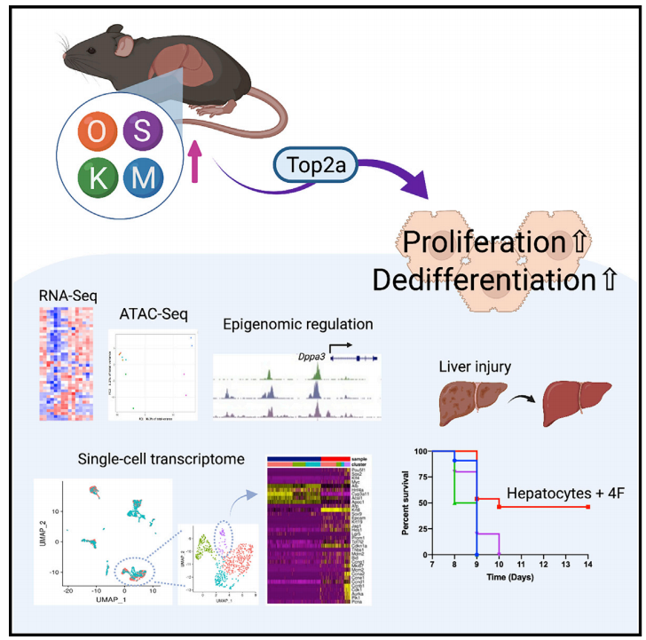
Hepatocyte-specific 4F expression induces cell proliferation and dedifferentiation
Hepatocyte-specific 4F expression induces a global change in DNA accessibility
Top2a is required for cellular reprogramming in vitro and in vivo
In vivo reprogramming has beneficial effects on regenerative capacity
Authors
Tomoaki Hishida, Mako Yamamoto,
Yuriko Hishida-Nozaki, ...,
Pradeep Reddy, Guang-Hui Liu,
Juan Carlos Izpisua Belmonte
Correspondence
该Email地址已收到反垃圾邮件插件保护。要显示它您需要在浏览器中启用JavaScript。
In brief
In regenerating animals, such as fish and salamanders, dedifferentiation followed by proliferation contributes to tissue regeneration. Hishida et al. show that hepatocyte-specific cellular reprogramming induces cell proliferation and dedifferentiation in the liver and enhances liver regenerative capacity through topoisomerase2-mediated partial reprogramming.
SUMMARY
Mammals have limited regenerative capacity, whereas some vertebrates, like fish and salamanders, are able to regenerate their organs efficiently. The regeneration in these species depends on cell dedifferentiation followed by proliferation. We generate a mouse model that enables the inducible expression of the four Yamanaka factors (Oct-3/4, Sox2, Klf4, and c-Myc, or 4F) specifically in hepatocytes. Transient in vivo 4F expression induces partial reprogramming of adult hepatocytes to a progenitor state and concomitantly increases cell proliferation. This is indicated by reduced expression of differentiated hepatic-lineage markers, an increase in markers of proliferation and chromatin modifiers, global changes in DNA accessibility, and an acquisition of liver stem and progenitor cell markers. Functionally, short-term expression of 4F enhances liver regenerative capacity through topoisomerase2-mediated partial reprogramming. Our results reveal that liver-specific 4F expression in vivo induces cellular plasticity and counteracts liver failure, suggesting that partial reprogramming may represent an avenue for enhancing tissue regeneration.

- 星期三, 28 8月 2024
The NADPARK study: A randomized phase I trial of nicotinamide riboside supplementation in Parkinson’s disease
SUMMARY
We conducted a double-blinded phase I clinical trial to establish whether nicotinamide adenine dinucleotide (NAD) replenishment therapy, via oral intake of nicotinamide riboside (NR), is safe, augments cerebral NAD levels, and impacts cerebral metabolism in Parkinson’s disease (PD). Thirty newly diagnosed, treatmentnaive patients received 1,000 mg NR or placebo for 30 days. NR treatment was well tolerated and led to a significant, but variable, increase in cerebral NAD levels—measured by 31phosphorous magnetic resonance spectroscopy—and related metabolites in the cerebrospinal fluid. NR recipients showing increased brain NAD levels exhibited altered cerebral metabolism, measured by 18fluoro-deoxyglucose positron emission to mography, and this was associated with mild clinical improvement. NR augmented the NAD metabolome and induced transcriptional upregulation of processes related to mitochondrial, lysosomal, and proteasomal function in blood cells and/or skeletal muscle. Furthermore, NR decreased the levels of inflammatory cytokines in serum and cerebrospinal fluid. Our findings nominate NR as a potential neuroprotective therapy for PD, warranting further investigation in larger trials.

- 星期二, 27 8月 2024
eIF5A hypusination, boosted by dietary spermidine, protects from premature brain aging and mitochondrial dysfunction
SUMMARY
Mitochondrial function declines during brain aging and is suspected to play a key role in age-induced cognitive decline and neurodegeneration. Supplementing levels of spermidine, a body-endogenousmetabolite, has been shown to promote mitochondrial respiration and delay aspects of brain aging. Spermidine serves as the aminobutyl group donor for the synthesis of hypusine (Nε -[4-amino-2-hydroxybutyl]-lysine) at a specific lysine residue of the eukaryotic translation initiation factor 5A (eIF5A). Here, we show that in the Drosophila brain, hypusinated eIF5A levels decline with age but can be boosted by dietary spermidine. Several genetic regimes of attenuating eIF5A hypusination all similarly affect brain mitochondrial respiration resembling age-typical mitochondrial decay and also provoke a premature aging of locomotion and memory formation in adult Drosophilae. eIF5A hypusination, conserved through all eukaryotes as an obviously critical effector of spermidine, might thus be an important diagnostic and therapeutic avenue in aspects of brain aging provoked by mitochondrial decline.

- 星期一, 26 8月 2024
Telomeres: history, health, and hallmarks of aging
Deepavali Chakravarti,1 Kyle A. LaBella,1 and Ronald A. DePinho1,*
1Department of Cancer Biology, The University of Texas MD Anderson Cancer Center, Houston, TX 77030, USA
*Correspondence: 该Email地址已收到反垃圾邮件插件保护。要显示它您需要在浏览器中启用JavaScript。
https://doi.org/10.1016/j.cell.2020.12.028
SUMMARY
The escalating social and economic burden of an aging world population has placed aging research at center stage. The hallmarks of aging comprise diverse molecular mechanisms and cellular systems that are interrelated and act in concert to drive the aging process. Here, through the lens of telomere biology, we examine how telomere dysfunction may amplify or drive molecular biological processes underlying each hallmark of aging and contribute to development of age-related diseases such as neurodegeneration and cancer. The intimate link of telomeres to aging hallmarks informs preventive and therapeutic interventions designed to attenuate aging itself and reduce the incidence of age-associated diseases.
