文献精选


This article is excerpted from the《Frontiers in Endocrinology》by Wound World
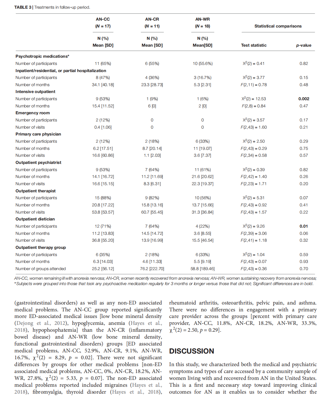
This article is excerpted from the《Frontiers in Psychology》by Wound World

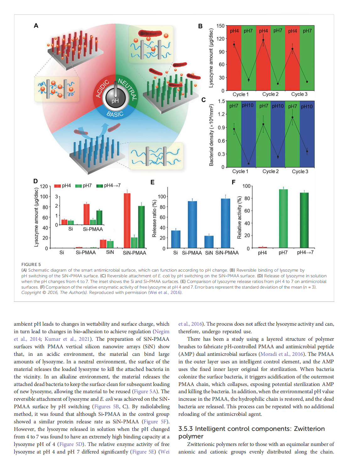
This article is excerpted from the《Frontiers in Bioengineering and Biotechnology》by Wound World

This article is excerpted from the《Frontiers in Surgery》by Wound World
